Monthly Archives: October 2025

Creativity keeps the mind young, says worldwide study on brain health

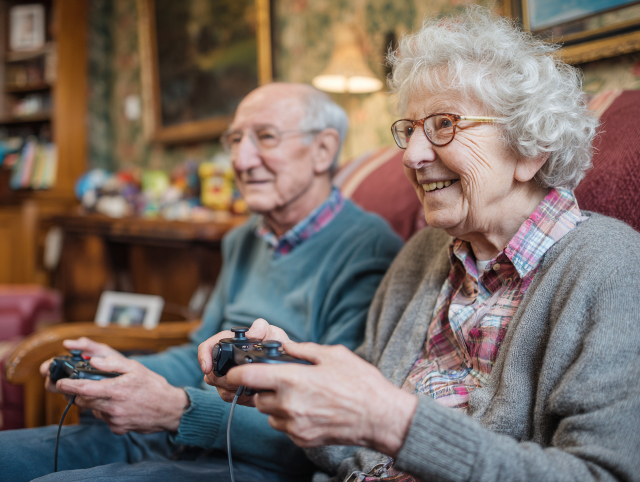
Engaging in creative experiences such as music, dance, visual arts, and even specially designed video games can significantly slow the process of brain ageing while promoting overall cognitive vitality, according to a groundbreaking international study recently published in Nature Communications. While creativity has long been celebrated for its aesthetic, emotional, and cultural value, this research provides the first large-scale empirical evidence that creative engagement has a measurable protective effect on brain health. By integrating neuroscientific techniques with behavioural data, the study opens a new frontier in understanding how the brain responds to artistic and imaginative pursuits, suggesting that creativity may be as essential to a healthy mind as diet and exercise are to a healthy body.

The study, conducted across 13 countries and involving more than 1,400 participants, included individuals with varying degrees of creative expertise—ranging from professional tango dancers, musicians, and visual artists to hobbyists, learners, and non-experts. Using advanced brain imaging techniques such as electroencephalography (EEG) and magnetoencephalography (MEG), researchers assessed biological versus chronological brain age through “brain clock” models. These models, which estimate the biological age of the brain based on patterns of neural activity, revealed that those regularly involved in creative activities exhibited “younger” brain profiles. Even participants who engaged in short-term creative training, such as brief video game sessions, showed measurable—albeit smaller—cognitive and neurological benefits, underscoring that creativity’s advantages are accessible to everyone, regardless of skill level.

Dr Agustin Ibanez, senior and corresponding author of the study, Professor of Brain Health at the Global Brain Health Institute (GBHI) and School of Medicine, Trinity College Dublin, and Director of the Latin American Brain Health Institute (BrainLat) at Universidad Adolfo Ibáñez, emphasised the transformative implications of the findings. “Creativity emerges as a powerful determinant of brain health, comparable to exercise or diet,” he said. “Our results open new avenues for creativity-based interventions to protect the brain against ageing and disease. Our study also showed that brain clocks can be used to monitor interventions aimed to improve brain health.” Echoing this sentiment, Dr Carlos Coronel, the study’s first author and a postdoctoral fellow at the GBHI, highlighted the accessibility of creative engagement: “You do not need to be an expert to benefit from creativity. Indeed, we found that learners gained from brief video game training sessions.”

A central feature of the research is its innovative use of brain clocks as diagnostic and predictive tools. Traditionally, brain clocks have been employed to measure negative influences on brain health, such as genetic predispositions, exposure to pollution, and socioeconomic stressors. This study, however, marks one of the first times these models have been used to capture positive influences—demonstrating that creativity can actively preserve and rejuvenate neural integrity. The researchers discovered that creative engagement not only correlates with youthful brain profiles but also strengthens functional connectivity in key regions vulnerable to neurodegeneration, such as the hippocampus, prefrontal cortex, and parietal lobes. Computational modelling further revealed that creative activities enhance the efficiency of brain networks, improving information processing and adaptability—mechanisms believed to underlie creativity’s neuroprotective power.

Beyond its neuroscientific significance, the study carries profound implications for public health and social policy. By illustrating that creativity can act as a low-cost, accessible means of promoting brain resilience, the findings advocate for a broader, more holistic conception of healthy ageing. As Dr Ibanez observed, “This is not relevant only for neuroscience, but it is a cultural and policy opportunity. Our societies need to reimagine healthy ageing through both medical and non-medical domains, such as creativity, arts, and play.” The authors propose that creative engagement could eventually be prescribed alongside exercise and dietary interventions as part of preventive healthcare strategies, particularly for populations at risk of cognitive decline. Such an approach would represent a significant paradigm shift, positioning creativity as a fundamental component of lifelong brain health rather than a recreational luxury.

Dr Coronel added that the diversity of creative experiences examined—ranging from tango dancing to video gaming—illustrates the universality of creativity’s benefits. “While these creative experiences differ, they share a common thread: they help protect brain connections that are vulnerable to accelerated ageing,” he said. “Because everyone’s hobbies and interests are unique, it’s encouraging to know there are many ways to support brain health through creativity.” In essence, the study underscores that creativity, in all its forms, offers a powerful form of cognitive nourishment. It affirms that engaging the imagination, expressing oneself artistically, or mastering a new skill does more than enrich life emotionally and culturally—it literally keeps the brain younger, healthier, and more adaptable. The research thus not only transforms our understanding of creativity’s role in the ageing process but also redefines it as a cornerstone of preventive neuroscience and a vital tool in the pursuit of longevity and mental well-being.

More information: Carlos Coronel et al, Creative experiences and brain clocks, Nature Communications. DOI: 10.1038/s41467-025-64173-9

Journal information: Nature Communications Provided by Trinity College Dublin

Calcium supplements not tied to dementia risk, ECU research shows

New research from Edith Cowan University (ECU), Curtin University and the University of Western Australia has found no evidence that calcium supplements increase the long-term risk of dementia, easing earlier concerns about their possible impact on brain health in older women. The study helps clarify years of uncertainty surrounding calcium monotherapy, which is commonly used to support bone health in later life.

The investigation drew on data from a previous five-year clinical trial involving 1,460 older women who received either calcium supplements or a placebo. Over a follow-up period of more than 14 years, researchers found that taking calcium did not raise the likelihood of developing dementia. This finding reassures both patients and healthcare providers that calcium supplementation remains safe in the context of cognitive decline.

ECU PhD student Ms Negar Ghasemifard explained that calcium supplements are widely recommended to prevent or manage osteoporosis, a condition affecting roughly one in five women over the age of seventy. She noted that previous studies had raised concerns about a potential link between calcium and dementia risk. Still, these new results provide confidence in the continued use of calcium supplements for bone protection without fear of cognitive harm.

Senior Research Fellow Dr Marc Sim emphasised that the study’s strength lies in its design. By adjusting for supplement compliance, dietary intake, lifestyle factors, and genetic risk, the researchers confirmed that their findings remained consistent. Unlike earlier observational studies that could not rule out hidden confounding factors, this double-masked, placebo-controlled randomised clinical trial provided more reliable evidence.

Dr Sim added that the trial’s long duration and careful tracking of dosage and compliance strengthened the credibility of the results. With 730 participants receiving calcium and 730 receiving placebo over five years, followed by a median of 14.5 years of monitoring, the data indicated no significant link between calcium use and all-cause dementia risk in older women.

While the findings are encouraging, ECU’s Centre for Precision Health Director Professor Simon Laws cautioned that further research is needed to determine whether similar results would apply to other groups, such as men or younger women who begin supplementation earlier in life. He suggested that future trials should include detailed brain health assessments as primary outcomes to confirm the long-term cognitive safety of calcium supplements.

More information: Negar Ghasemifard et al, Calcium supplementation and the risk of dementia in the Perth Longitudinal Study of Aging Women: a post-hoc analysis of a randomised clinical trial for fracture prevention, The Lancet. DOI: 10.1016/j.lanwpc.2025.101694

Journal information: The Lancet Provided by Edith Cowan University

Researchers Rewind Alzheimer’s in Mice with Nanoparticle Breakthrough

A research team co-led by the Institute for Bioengineering of Catalonia (IBEC) and the West China Hospital of Sichuan University (WCHSU), in partnership with collaborators in the United Kingdom, has developed a groundbreaking nanotechnology approach that reverses Alzheimer’s disease in mice. Unlike conventional nanomedicine, which typically uses nanoparticles as passive carriers for drugs, this method employs “supramolecular drugs”—nanoparticles that are themselves biologically active. Instead of targeting neurons directly, the therapy focuses on repairing the blood–brain barrier (BBB), the protective interface that regulates what enters and leaves the brain. By restoring the BBB’s natural function, the team achieved a reversal of Alzheimer’s pathology in animal models, offering a transformative new direction for neurodegenerative disease treatment.

The brain is one of the body’s most energy-hungry organs, consuming about 20 per cent of an adult’s energy and up to 60 per cent in children. This energy is delivered through a vast and intricate network of around one billion capillaries, ensuring that each neuron is adequately nourished. The study underscores that vascular health is central to brain function: when the brain’s blood vessels deteriorate, cognitive decline often follows. This finding highlights that Alzheimer’s is not solely a neuronal disorder but also a disease of the brain’s vasculature. Repairing this vascular system, therefore, not only halts progression but can also help restore lost brain function.

The blood–brain barrier plays a vital defensive role, shielding the brain from toxins and pathogens. In Alzheimer’s disease, however, this barrier becomes compromised, trapping toxic “waste proteins” such as amyloid-β (Aβ) inside the brain. The researchers discovered that their supramolecular nanoparticles could reset this clearance system, allowing Aβ to pass safely into the bloodstream and be removed. In mice genetically engineered to overproduce Aβ and mimic Alzheimer’s symptoms, three doses of the nanoparticle treatment led to a 50 to 60 per cent reduction in Aβ levels within just one hour. According to Junyang Chen, co-author of the study and researcher at WCHSU, this rapid effect marked a breakthrough in addressing the disease’s root cause rather than merely managing symptoms.

The behavioural results were equally impressive. Treated mice displayed marked improvements in memory and cognitive function, even at advanced stages of the disease. In one key experiment, a 12-month-old mouse—the equivalent of a 60-year-old human—received the nanoparticle treatment and was observed for six months. By the end of the study, the now 18-month-old mouse (roughly equivalent to a 90-year-old human) had recovered behaviour comparable to a healthy animal. “The long-term effect comes from restoring the brain’s vasculature,” explained Professor Giuseppe Battaglia, ICREA Research Professor at IBEC and leader of the Molecular Bionics Group. “Once the vascular system begins to function again, it clears amyloid-beta and other harmful molecules, allowing the brain to regain its natural balance.”

The key to this recovery lies in restoring amyloid-β clearance through the receptor LRP1, which typically acts as a molecular gatekeeper. In Alzheimer’s, excessive Aβ overwhelms this receptor, halting the brain’s ability to remove waste proteins. The supramolecular nanoparticles act as molecular “switches,” mimicking the natural ligands of LRP1 to reactivate the clearance process. Designed with molecular precision, these nanoparticles are built from the bottom up, featuring carefully engineered size and surface ligands that allow them to interact specifically with cell receptors. This precision not only enables the removal of toxic proteins but also reinstates vascular health, reducing inflammation and improving overall brain homeostasis.

This study redefines how Alzheimer’s disease can be treated by addressing its vascular origins rather than focusing solely on damaged neurons. By repairing the blood–brain barrier and restoring the brain’s natural waste-clearing mechanisms, the supramolecular drugs demonstrated an ability to reverse cognitive decline in ways previously thought impossible. As Lorena Ruiz Pérez, researcher at IBEC’s Molecular Bionics Group and Serra Hunter Assistant Professor at the University of Barcelona, concluded, “Our study demonstrated remarkable efficacy in achieving rapid amyloid-β clearance, restoring healthy blood–brain barrier function, and leading to a striking reversal of Alzheimer’s pathology.” This innovative therapy represents a significant leap forward in the fight against dementia and opens new horizons for nanomedicine’s role in combating neurodegenerative disease.

More information: Junyang Chen et al, Rapid amyloid-β clearance and cognitive recovery through multivalent modulation of blood–brain barrier transport, Signal Transduction and Targeted Therapy. DOI: 10.1038/s41392-025-02426-1

Journal information: Signal Transduction and Targeted Therapy Provided by Institute for Bioengineering of Catalonia (IBEC)

Comprehensive protein network reveals how neural communication falters in Alzheimer’s disease

A pioneering investigation from the Icahn School of Medicine at Mount Sinai has illuminated one of the most comprehensive portraits yet of how brain cells communicate—and how that intricate dialogue deteriorates in Alzheimer’s disease. The research, published in Cell on 25 September, represents a major leap in understanding the cellular crosstalk underlying neurodegeneration. By mapping the complex web of protein interactions that govern communication between neurons and supporting cells, the team has revealed a systemic breakdown in this network that could explain why brain function collapses as the disease progresses. Notably, the study not only highlights how these failures occur but also identifies several potential therapeutic targets that may help restore healthy cellular communication and slow cognitive decline.

The Mount Sinai-led team analysed protein activity in post-mortem brain tissue samples from nearly two hundred individuals, both with and without Alzheimer’s disease. Using cutting-edge proteomics—the large-scale study of proteins—and advanced computational modelling, the researchers created an extensive “map” of how more than 12,000 proteins interact within the brain. This holistic approach allowed them to detect patterns of dysfunction that traditional methods, which tend to focus on single molecules such as amyloid or tau, often overlook. Their findings revealed that disruptions in communication between neurons and glial cells—particularly astrocytes and microglia—are central to the pathology of Alzheimer’s. These glial cells, which generally provide essential support, protection, and maintenance for neurons, appear to lose their regulatory balance in the disease, becoming hyperactive and contributing to inflammation and neural damage.

The study’s senior author, Bin Zhang, PhD, Willard T.C. Johnson Research Professor of Neurogenetics and Director of the Center for Transformative Disease Modeling, emphasised that Alzheimer’s must be understood as more than an accumulation of plaques or dying neurons. “Our work shows that the breakdown of communication within the brain’s ecosystem—between neurons and glial cells—is likely a key factor driving disease progression,” he explained. This interpretation challenges decades of Alzheimer’s research, which the amyloid hypothesis has dominated. While amyloid plaques and tau tangles are indeed hallmark features of the condition, targeting them directly has yielded only limited success in clinical trials. The Mount Sinai study suggests that a broader systems-level failure may be the actual cause, where the delicate equilibrium of cellular interactions is disrupted long before symptoms appear.

A particularly striking aspect of the research is the identification of a protein called AHNAK as a major driver of pathological changes in the Alzheimer’s brain. AHNAK, which is abundant in astrocytes, was found to increase significantly as the disease advanced. Its levels correlated with heightened concentrations of toxic proteins such as amyloid beta and tau. Laboratory experiments using human stem-cell-derived brain models confirmed that reducing AHNAK expression led to decreased tau accumulation and improved neuronal function. Co-senior author Dongming Cai, MD, PhD, of the University of Minnesota, noted that these results “suggest that AHNAK could be a promising therapeutic target. By lowering its activity, we saw both less toxicity and more neuronal vitality.” This discovery provides compelling evidence that modulating glial activity could help rebalance brain function, potentially reversing or slowing the degenerative process.

The study’s use of an “unsupervised” analytic framework was another major innovation. Co-senior author Junmin Peng, PhD, from St. Jude Children’s Research Hospital, explained that the approach allowed researchers to identify critical protein networks without bias. “By examining how thousands of proteins interact, rather than starting with assumptions about which ones matter, we gained an unprecedented view of the proteomic alterations underlying Alzheimer’s,” he said. This systems-biology perspective enabled the discovery of over three hundred previously underexplored proteins linked to the disease. These proteins may hold the key to understanding individual variations in disease onset and progression, offering new directions for both basic research and clinical investigation.

Another important insight from the study concerns how genetic and biological factors modulate these protein networks. Individuals carrying the APOE4 gene variant—a well-known risk factor for Alzheimer’s—showed markedly different patterns of network disruption compared with those without the gene. Similarly, the data suggested potential differences between men and women in how these cellular communication systems respond to stress and disease. Such findings underscore the importance of personalised medicine approaches in future Alzheimer’s research, recognising that therapeutic strategies may need to be tailored to a patient’s unique genetic and molecular background. The team has made their entire dataset publicly available, inviting scientists worldwide to explore these connections and accelerate progress across the field.

Ultimately, this study redefines Alzheimer’s as a disorder of broken communication rather than merely one of toxic buildup. By revealing how the interplay between neurons and glia governs the brain’s overall resilience, it opens a new conceptual framework for understanding neurodegeneration. As Dr Zhang eloquently summarised, “This research helps us see Alzheimer’s not as a static disease but as a dynamic failure of interaction. If we can understand where those conversations between brain cells go wrong, we can start finding ways to repair them.” The work thus marks a significant step towards developing therapies that target the root causes of cognitive decline—by restoring balance and harmony to the brain’s intricate network of cellular communication rather than simply clearing away its debris.

More information: Bin Zhang et al, Multiscale proteomic modeling reveals protein networks driving Alzheimer’s disease pathogenesis, Cell. DOI: 10.1016/j.cell.2025.08.038

Journal information: Cell Provided by The Mount Sinai Hospital / Mount Sinai School of Medicine

The Real Priorities of Older People Might Surprise You

A recent study from Case Western Reserve University has revealed a surprising insight into the lives and priorities of senior citizens receiving treatment at walk-in care clinics across the United States. While many might assume that health would dominate their concerns, the research found otherwise. Although maintaining health remains a significant priority, older adults rated “social activities and inclusiveness” as the most important aspect of their lives. This discovery challenges the common perception that medical or physical well-being automatically tops the list of older adults’ values. Conducted on a national scale, the study sheds light on the complexity of what truly matters to individuals aged sixty-five and above when they seek care at these outpatient facilities.

The research team analysed responses from roughly 388,000 patients who visited walk-in ambulatory care clinics between January 2021 and March 2024. These clinics, often located in neighbourhood pharmacies or community health centres, provide medical services for minor illnesses and injuries without requiring an appointment. Patients were asked a simple but revealing open-ended question: “What matters most to you?” The responses offered a profound glimpse into the mindset of ageing Americans. Nearly half of the participants identified social engagement—interactions, group activities, and feeling included within a community—as their top concern. Health followed at 21%, while independence and family togetherness ranked next at 17% and 10.5% respectively. Such figures point to a deeply human truth: older adults crave belonging and connection as much as, if not more than, they value physical well-being.

The study was co-led by Nicholas Schiltz, an assistant professor at Case Western Reserve University’s Frances Payne Bolton School of Nursing. Schiltz admitted that the findings came as a surprise, particularly given that the data were gathered from patients in acute-care settings. “I would have thought health would have been a little bit higher since this is an acute-care setting,” he explained, suggesting that the outcome reveals a broader truth about the emotional and social dimensions of ageing. Schiltz emphasised that older adults, like people of all ages, are multifaceted individuals whose lives cannot be reduced to a single factor such as physical health. Their identities and sense of purpose remain closely tied to community participation, independence, and family bonds.

The researchers designed the study to deepen understanding of patients’ priorities with the ultimate goal of improving health outcomes and personalising care. By identifying what matters most to older adults, clinicians can create more meaningful care plans that align with their values and lifestyles. For instance, if a patient values independence or social engagement above all else, care providers might adjust medication plans to reduce the risk of dizziness or falls that could limit mobility. They might also recommend physical therapy or assistive devices that enable patients to continue participating in community activities. In this way, healthcare becomes not merely a matter of treatment, but a partnership that respects the patient’s individuality and broader quality of life.

The study’s demographic data adds further depth to its findings. Among the respondents, nearly two-thirds were women, three-quarters identified as white, 5% as Black, 4.7% as Hispanic, and 2.7% as Asian. Interestingly, the researchers found that these differences in background had little bearing on what participants valued most. Across all races, sexes, and ethnic groups, social belonging remained the dominant concern. This universality underscores that the desire for inclusion and meaningful relationships transcends demographic boundaries. Whether in urban centres or rural towns, whether affluent or working-class, older adults across the country share a strikingly similar vision of what gives their lives purpose.

Mary Dolansky, the Sarah C. Hirsh Professor at the Frances Payne Bolton School of Nursing and co-leader of the study, placed the research within a broader national movement toward age-friendly health systems. She noted that the findings align with the mission of The John A. Hartford Foundation’s Age-Friendly Health Systems initiative, which aims to ensure that all older adults receive evidence-based, holistic care. Dolansky highlighted that effective elderly care must extend beyond physical treatment to encompass safe medication practices, assessments of mental health, cognitive function, and mobility. With the population ageing rapidly, she stressed the urgency of adapting the healthcare system to meet these evolving needs. The Case Western study thus serves not only as an academic contribution but as a call to action: to view older adults not just as patients with medical conditions, but as individuals with complex emotional, social, and familial lives deserving of dignity and understanding.

More information: Nicholas Schiltz et al, Patient-Centered Priorities for Older Adults in Ambulatory Care, JAMA Network Open. DOI: 10.1001/jamanetworkopen.2025.35769

Journal information: JAMA Network Open Provided by Case Western Reserve University

Little worms, mighty memories: decoding the brain’s secrets

In a striking discovery that challenges conventional wisdom about memory, researchers at Flinders University have found that forgetting is not merely a flaw in the brain’s function but an essential, carefully regulated process. Their work shows that dopamine, the chemical often associated with learning, motivation, and reward, also plays a crucial role in helping the brain forget. Far from being a failure, forgetting serves a vital purpose in maintaining cognitive flexibility, allowing the brain to stay focused and adaptable rather than overwhelmed by an overload of information.

Led by neuroscientist Dr Yee Lian Chew and PhD student Anna McMillen from the Flinders Health and Medical Research Institute (FHMRI), the study was published in the Journal of Neurochemistry. It used tiny worms known as Caenorhabditis elegans. Though each worm measures just a millimetre long and has only 300 neurons, it shares about 80 per cent of its genes with humans, making it an ideal model for exploring brain mechanisms. The team trained these worms to associate a scent with food and then observed how long they retained that memory. Remarkably, worms that lacked dopamine held onto their memories much longer, suggesting that dopamine is the trigger that allows the brain to let go.

Dr Chew explains that this ability to forget is vital to a healthy mind. “We often think of forgetting as a failure,” she says, “but it’s actually essential. If we remembered everything, our brains would be overwhelmed. Forgetting helps us stay focused and flexible.” Further investigation revealed that two specific dopamine receptors — DOP-2 and DOP-3 — work together to control forgetting. When both receptors were disabled, the worms could no longer forget, mirroring the behaviour of dopamine-deficient ones. Even partial restoration of dopamine did not help; the entire system needed to function correctly for normal forgetting to resume.

The researchers believe this discovery has wide-ranging implications for human health. Since dopamine also plays a significant role in neurological conditions such as Parkinson’s disease, understanding how it regulates memory loss could pave the way for new treatments. “By studying the worm brain, we can understand the same chemical processes that happen in humans,” Dr Chew explains. “This could help us learn why memory changes with age and in diseases where dopamine is disrupted.” Their goal is to pinpoint how dopamine acts on specific neurons to erase outdated memories while preserving those that are relevant selectively.

The findings build on earlier studies in fruit flies, suggesting that dopamine-driven forgetting is a universal biological mechanism shared across species. This insight reframes forgetting as an active, purposeful part of brain function rather than a defect. As Dr Chew concludes, “It’s exciting to see that something so fundamental is shared across species. It means we’re uncovering a deep biological truth that could one day lead to breakthroughs in understanding human memory.” Through the humble worm, researchers are revealing that forgetting — often seen as loss — may in fact be one of the brain’s greatest strengths.

More information: Yee Lian Chew et al, Dopaminergic Modulation of Short-Term Associative Memory in Caenorhabditis elegans, Journal of Neurochemistry. DOI: 10.1111/jnc.70200

Journal information: Journal of Neurochemistry Provided by Flinders University

Arts participation linked to lower rates of heart disease, diabetes, and other major causes of death, large-scale study reveals

Art is far more than an indulgence for the imagination or a vehicle for creative self-expression; it can also serve as a potent and practical instrument for public health. That is the central message emerging from a landmark international study commissioned by the Jameel Arts and Health Lab, which scrutinised nearly one hundred research projects spanning twenty-seven countries. The study explored how participation in creative and cultural activities—ranging from music, dance, theatre, and storytelling to painting, crafts, and gardening—can contribute to the prevention of some of the world’s deadliest non-communicable diseases, including heart disease, diabetes, and cancer. These conditions, which account for an estimated seventy-four per cent of preventable deaths worldwide, represent an immense burden on healthcare systems and societies. The findings suggest that by embracing artistic engagement as a component of public health strategies, communities could reduce risk factors for disease while enhancing quality of life and well-being.

The timing of this research is particularly significant, as it coincides with an international meeting convened by the World Health Organization devoted to the prevention of non-communicable diseases. As policymakers, clinicians, and researchers seek new ways to reduce the global prevalence of chronic illnesses, the study offers compelling evidence that the arts can play a transformative role in shaping healthier societies. Rather than existing on the margins of healthcare policy, the arts are presented here as integral to public health infrastructure. Through active participation in creative programmes, individuals may not only improve their physical and mental health but also strengthen social bonds and community resilience. This growing body of evidence invites a reimagining of health promotion—one that values creativity, emotion, and culture as vital components of human flourishing.

At the heart of the study’s message is a call to shift the focus of healthcare from treatment to prevention. Jill Sonke, PhD, the director of research initiatives at the University of Florida’s Centre for Arts in Medicine and the lead author of the study, argued that the arts have untapped potential in preventing chronic disease before it develops. “We don’t want just to treat these diseases; we want to prevent them,” Sonke remarked. She emphasised that investments in health should “move upstream” from clinical treatment toward prevention, with creative engagement serving as a central pillar in that effort. The study she co-led—published on 18 September in Nature Medicine—represents one of the most comprehensive analyses of its kind, incorporating data from ninety-five studies and more than 230,000 participants. Supported in part by the State of Florida Division of Arts and Culture and New York University, the research provides a scientifically grounded framework for integrating artistic practice into health policy.

Working alongside Sonke was Michael Tan, PhD, dean of research and knowledge exchange at the University of the Arts Singapore, who helped direct the international team of scholars involved in the project. Their collective findings demonstrate that participation in the arts can yield measurable benefits across a variety of health indicators. Meanwhile, co-author Nisha Sajnani, PhD, a professor at NYU Steinhardt and co-director of the Jameel Arts and Health Lab, underscored the importance of treating the arts not as a luxury, but as essential public health infrastructure. “If we are serious about reducing the global burden of non-communicable diseases, we must treat the arts as essential to public health infrastructure,” she noted. Sajnani highlighted that artistic and cultural programmes offer cost-effective, scalable tools for prevention that can be embedded within community health initiatives. By grounding these programmes in partnership with local communities, she argued, they can bridge equity gaps, increase access, and promote the adoption of healthier behaviours.

One of the study’s most striking conclusions is that arts-based programmes make health education more engaging, memorable, and effective. For example, a community play focused on healthy eating can translate nutritional guidance into relatable narratives; a dance class can encourage physical activity without the intimidation of gym environments; and a community gardening project can nurture both physical well-being and social connectedness. These activities convert abstract health information into lived experience. Notably, the research suggests that the social and emotional connections fostered by such programmes are as vital to health outcomes as the behaviours themselves. Participation in group-based, creative initiatives tends to sustain engagement far longer than traditional health campaigns, which often struggle to maintain public interest. When people are motivated by joy, creativity, and shared purpose, they are more likely to adopt and sustain healthy habits.

Moreover, the cultural relevance of artistic interventions enhances their effectiveness in ways that traditional medical campaigns often overlook. Programmes that draw upon local cultural forms—such as indigenous storytelling, traditional dance, or regional crafts—resonate more deeply within their communities. They provide familiar contexts through which important health messages can be conveyed and normalised. In this way, the arts serve as a bridge between scientific knowledge and cultural practice, ensuring that prevention campaigns are not only informative but also inclusive and meaningful. The study found that when health messages are culturally grounded, participation rates increase, behavioural change becomes more sustainable, and public trust in health institutions improves. These insights illustrate how culturally responsive approaches to public health can create more equitable systems of care.

Despite the strength of its findings, the research team acknowledged that there is still much to learn about the long-term impacts of arts participation on health outcomes. Most of the existing studies have been conducted in high-income countries, which raises questions about how such interventions might function in lower-income or resource-limited settings. Additionally, few studies have tracked participants over extended periods to determine whether the health benefits of creative engagement persist. Nevertheless, the evidence so far points towards a powerful conclusion: that the arts are not a mere luxury or afterthought in healthcare, but a vital component of disease prevention. By weaving creativity into the fabric of community life, societies can build resilience, reduce the prevalence of chronic illness, and enrich human experience.

Ultimately, this international study invites a profound reconsideration of how we define and promote health. Medicine and art, often viewed as separate realms, may in fact be complementary forces working toward a common goal: the flourishing of human life. By engaging people’s emotions, imaginations, and cultural identities, the arts can transform abstract health directives into meaningful, lived realities. As governments and organisations seek new ways to confront the escalating global crisis of non-communicable diseases, these findings present a hopeful and holistic vision—one in which creativity itself becomes an essential part of the world’s public health toolkit.

More information: Jill Sonke et al, The arts for disease prevention and health promotion: a systematic review, Nature Medicine. DOI: 10.1038/s41591-025-03962-7

Journal information: Nature Medicine Provided by University of Florida

Back Pain Linked to Increased Risk of Common Illnesses

Back pain, often dismissed as a simple musculoskeletal complaint, is increasingly being recognised as a complex health issue that extends far beyond the spine. Recent research suggests that chronic back pain may serve as an early warning sign of other severe conditions, including cardiovascular disease, arthritis, diabetes, and even cancer. This emerging evidence challenges the conventional understanding of back pain as merely a localised problem, positioning it instead as a possible indicator of systemic ill health. In particular, the findings highlight that individuals who experience chronic back pain are at a considerably higher risk of developing a range of common and serious diseases than those without it. Such discoveries could reshape how clinicians and policymakers approach prevention and treatment, encouraging a more integrated model of healthcare that recognises the interconnection between pain and broader physiological decline.

The study underpinning these findings was led by Associate Professor Rafael Zambelli Pinto, a physiotherapist and researcher in musculoskeletal health at the University of Technology Sydney (UTS). It was published in the Brazilian Journal of Physical Therapy and conducted in collaboration with an international team from several universities, including the Universidade Federal de Minas Gerais, Universidade Estadual Paulista, and the University of São Paulo in Brazil, as well as the University of Sydney, UNSW, and NeuRA in Australia. The team utilised data from the 2019 National Health Survey in Brazil, encompassing nearly 90,000 participants, making it one of the most comprehensive investigations of its kind in a middle-income country. Approximately one in five respondents reported living with chronic back pain, a proportion strikingly similar to rates observed in higher-income nations, suggesting that this is a truly global public health issue that transcends geography, income, and lifestyle.

The results painted a striking picture of the health burden borne by individuals with chronic back pain. The researchers discovered that conditions such as cardiovascular disease, arthritis, and depression were far more prevalent among those with persistent pain. Diabetes, cancer, asthma, and chronic lung conditions also occurred at higher rates. Statistically, people with back pain were found to be 17 per cent more likely to report cardiovascular disease, 15 per cent more likely to suffer from arthritis, and 12 per cent more likely to experience clinical depression than their pain-free counterparts. This clustering of physical and mental health problems points to a deeper biological and psychosocial connection—one that suggests chronic pain is not an isolated ailment but part of a larger web of interrelated risk factors and bodily dysfunctions.

In Australia, chronic back pain represents a particularly significant burden. The Australian Institute of Health and Welfare estimates that around four million Australians are living with chronic back problems. Nearly three-quarters of these individuals, particularly those aged over forty-five, also suffer from at least one additional chronic health condition. Those who experience both back pain and another illness—particularly arthritis, depression, or cardiovascular disease—report substantially higher levels of disability and activity limitation. Indeed, adults with both back pain and arthritis are more than twice as likely to experience severe restrictions in daily activities compared to those without this combination of conditions. These statistics underscore not only the widespread prevalence of back pain but also the profound effect it has on individuals’ physical capacity, mental wellbeing, and economic productivity.

Dr Zambelli Pinto has emphasised that while further research is needed to clarify the precise nature of the link between chronic back pain and other diseases, there are several plausible explanations rooted in shared lifestyle and behavioural risk factors. “While more research is needed to understand the link between chronic back pain and other non-communicable diseases, they often share underlying risk factors such as physical inactivity, obesity, stress and poor sleep,” he explained. These factors may feed into a self-reinforcing cycle: chronic pain leads to reduced mobility, which contributes to weight gain and cardiovascular strain; stress and poor sleep exacerbate pain sensitivity and inflammation, which in turn hinder recovery. This interdependence highlights how addressing back pain effectively requires more than managing discomfort—it demands confronting the broader determinants of health, such as diet, exercise, mental wellbeing, and rest.

The study concludes with a clear call for healthcare reform. Back pain is one of the most common reasons people visit their GP, yet the research suggests that treatment often remains too narrowly focused on symptom relief. Dr Zambelli Pinto and his colleagues argue for a more holistic, interdisciplinary approach—one that not only alleviates pain but also identifies and manages associated chronic diseases. “It’s not just about the spine,” he said. “These patients are carrying a heavier overall health burden that affects their independence and quality of life.” Comprehensive care pathways that integrate physiotherapy, psychological support, and medical screening for comorbidities could drastically improve both patient outcomes and system efficiency. In essence, back pain should be understood not merely as a mechanical failure of the body, but as a window into the individual’s broader state of health—a visible signal of hidden struggles that, if addressed early and collaboratively, could prevent more serious conditions from taking root.

More information: Rafael Zambelli Pinto et al, Prevalence of non-communicable diseases, multimorbidity, and their impact on activity limitations among adults with chronic back pain: a national population-based study in a middle-income country, Brazilian Journal of Physical Therapy. DOI: 10.1016/j.bjpt.2025.101241

Journal information: Brazilian Journal of Physical Therapy Provided by University of Technology Sydney

Clues in urine: protein levels linked to dementia development

A new study led by researchers at Karolinska Institutet has found that people with higher levels of the protein albumin in their urine face an increased risk of developing dementia later in life. The findings, published in the Journal of Internal Medicine, point to a strong link between kidney health and brain function, challenging the long-held notion that dementia arises solely from processes within the brain. Although advancing age remains the most significant risk factor for dementia, scientists are increasingly recognising that conditions affecting other organs—particularly the kidneys—may contribute to the onset and progression of cognitive decline. This research highlights the importance of looking beyond the brain itself to understand and potentially prevent neurodegenerative diseases.

Albumin is a key protein in the blood that helps maintain fluid balance and transport hormones, vitamins, and enzymes throughout the body. Under normal conditions, albumin should not pass through the kidneys into the urine in significant quantities. When it does, the condition is known as albuminuria, an early sign that the kidneys’ filtering units, or glomeruli, are damaged. In this new study, researchers analysed data from 130,000 older adults living in Stockholm, all aged 65 or above and free from dementia when the study began. Over a follow-up period of about four years, seven per cent of participants went on to develop some form of dementia, allowing researchers to examine how urinary albumin levels correlated with cognitive decline.

The results were clear and compelling. Individuals with moderate albumin levels—between 30 and 299 milligrams per gram of creatinine—had a 25 per cent greater risk of developing dementia compared to those with normal levels, defined as less than 30 milligrams per gram. The risk was even higher for those with severe albuminuria, at or above 300 milligrams per gram, who faced a 37 per cent higher likelihood of developing dementia. This pattern held even after adjusting for other variables, including kidney function, blood pressure, and cardiovascular health. Such findings suggest that albuminuria is not merely a by-product of poor health but a potential independent marker of dementia risk, particularly for forms associated with vascular damage.

Indeed, the association was strongest for vascular dementia—the second most common type of dementia after Alzheimer’s disease—as well as for mixed dementia, which combines features of both vascular and Alzheimer’s pathology. Vascular dementia occurs when blood flow to the brain is compromised, typically due to conditions such as hypertension, diabetes, or stroke. The researchers propose that albuminuria may reflect systemic vascular dysfunction affecting both the kidneys and the brain. Because both organs depend on networks of fine blood vessels, damage to these delicate structures in one organ often mirrors similar processes in the other. As the study’s senior author, Assistant Professor Hong Xu of Karolinska Institutet’s Department of Neurobiology, Care Sciences and Society, explained, “The kidneys and the brain may seem like very different organs, but they share an important characteristic: both depend on a delicate network of small blood vessels. When the blood vessels in the kidneys are damaged, the same process often occurs in the brain.”

This interconnection between kidney and brain health extends to the blood–brain barrier, a semipermeable layer that protects neural tissue from harmful substances in the bloodstream. When this barrier is compromised, toxins and inflammatory molecules can penetrate the brain, leading to damage, inflammation, and the build-up of harmful proteins such as amyloid-beta and tau, which are closely linked to dementia. The study draws a parallel between this process and another. What happens in the kidneys: just as a damaged renal filter leaks proteins into the urine, a weakened blood–brain barrier allows unwanted materials into brain tissue. Over time, these cumulative effects can accelerate the deterioration of neural pathways and contribute to cognitive decline.

The implications of these findings are significant, suggesting that a simple urine test could become a valuable tool for assessing dementia risk. Routine screening for albuminuria is already standard practice in patients with diabetes and kidney disease, but this research broadens its potential use to include early dementia risk assessment. “These results underscore the importance of routine screening for albuminuria as part of early dementia risk assessment, especially in patients with high blood pressure, diabetes, cardiovascular disease, or kidney disease. Early detection of albuminuria could potentially delay or prevent the onset of dementia,” said Hong Xu. Such preventive strategies could enable clinicians to identify at-risk individuals long before cognitive symptoms emerge, allowing for early lifestyle or medical interventions.

Moreover, this study reinforces the importance of a holistic approach to dementia prevention. Rather than viewing dementia solely as a neurological disorder, it calls for an integrated model of care that addresses systemic health, particularly cardiovascular and renal function. Managing blood pressure, controlling blood sugar, reducing cholesterol, and maintaining vascular health may protect not only the heart and kidneys but also the brain. Lifestyle factors such as diet, exercise, and stress reduction can further help maintain healthy blood vessels throughout the body. The message is clear: protecting the brain begins with protecting the systems that sustain it.

Ultimately, the Karolinska Institutet study represents a significant step forward in understanding the biological bridges between the kidneys and the brain. By identifying albuminuria as a measurable, early marker of dementia risk, it offers clinicians and researchers a new pathway for early diagnosis and intervention. As the global population ages and dementia cases continue to rise, such discoveries are vital. They remind us that the body operates as an interconnected whole and that diseases once considered localised—such as kidney dysfunction—may, in fact, have far-reaching consequences for cognitive health. In essence, the research shows that what we see in the urine may hold vital clues to what is happening in the brain.

More information: Hong Xu et al, Albuminuria is associated with increased risk of dementia, independent of eGFR: The SCREAM project, Journal of Internal Medicine. DOI: 10.1111/joim.70022

Journal information: Journal of Internal Medicine Provided by Karolinska Institutet

Long-term human connections might delay the body’s natural ageing process

The cumulative influence of social advantages throughout one’s life — beginning with parental warmth in early childhood and extending through friendships, community engagement, and religious or spiritual participation in adulthood — appears to slow down the biological processes of ageing. Scientists have found that these enduring social benefits can effectively reset what are known as epigenetic clocks, which estimate a person’s biological age by examining chemical patterns on their DNA, specifically through a process called DNA methylation. In practical terms, this means that people who have experienced richer and more supportive social relationships across their lives tend to be biologically younger than their chronological years would suggest. Such findings reinforce the notion that the social bonds we cultivate may have profound, measurable effects not only on emotional well-being but on the very pace at which our bodies age.

This conclusion was drawn from research published in the October issue of Brain, Behavior and Immunity – Health, based on a detailed analysis of data from more than 2,100 adults who participated in the long-running Midlife in the United States (MIDUS) study. The research team, led by Professor Anthony Ong of Cornell University, examined how lifelong social support contributes to biological resilience. Their results showed that individuals who possessed higher levels of what they termed “cumulative social advantage” exhibited slower rates of epigenetic ageing and lower levels of chronic inflammation. The study presents compelling evidence that social connection operates as a biological buffer, reinforcing the body’s defences against the cellular wear and tear that accumulates with age.

At the heart of the study were two particular epigenetic clocks—known as GrimAge and DunedinPACE—which scientists consider among the most reliable predictors of morbidity and mortality. Ong and his colleagues found that participants with stronger, more stable networks of family, friends, and community ties had significantly younger biological profiles according to both measures. These results suggest that social connection can influence molecular processes linked to longevity, implying that the web of relationships people build over time can have a tangible, physical impact on the human body. In other words, nurturing relationships may not only enrich one’s emotional life but also alter biological systems in ways that slow ageing itself.

Ong described “cumulative social advantage” as a multidimensional concept encompassing the depth and diversity of one’s social experiences across life. The researchers identified four primary sources of social benefit: the warmth and emotional support received from parents during childhood; one’s sense of belonging within their community and neighbourhood; participation in religious or faith-based organisations; and continued emotional support from family and friends during adulthood. Together, these sources form a web of interconnected experiences that sustain psychological stability and physiological balance. They shape a person’s stress response, strengthen their immune system, and promote biological resilience — all of which are crucial in determining how rapidly or slowly the body ages.

The study also explored how sustained social advantage might influence the body’s regulatory systems — particularly those governing inflammation, gene expression, and hormonal balance. The findings showed that participants with higher social advantage had markedly lower levels of interleukin-6, a pro-inflammatory molecule that has been strongly associated with conditions such as cardiovascular disease, diabetes, and neurodegenerative disorders. Interestingly, there was no significant link between social advantage and short-term stress indicators such as cortisol or catecholamines. This suggests that the protective effects of social relationships may emerge from long-term physiological stability rather than temporary relief from stress. In essence, meaningful relationships appear to exert a steady, cumulative influence that preserves the body’s internal equilibrium.

Unlike earlier research that focused narrowly on specific social factors — such as whether a person is married or the number of friends they have — Ong’s study conceptualised “cumulative social advantage” as a layered and evolving phenomenon. It acknowledges that relationships are not static and that social capital grows, shifts, and deepens over time. Those who experience nurturing family bonds early in life are more likely to sustain and expand their social resources in adulthood, compounding their benefits across the decades. As Ong explained, “It’s not just about whether you have supportive friends now, but how your connections have grown and deepened over time. That accumulation shapes your health in ways we can measure biologically.” His words highlight a crucial point: social well-being is not built overnight but cultivated gradually through consistent emotional investment.

The broader implications of this research are both scientific and deeply human. It reframes ageing not merely as a biological inevitability governed by genetics or lifestyle choices but as a social process profoundly influenced by the quality and continuity of our relationships. A single act of friendship or a brief period of community involvement may not reverse cellular ageing. Still, the sustained presence of supportive relationships over many years can meaningfully slow the biological clock. Ong captures this idea with a vivid metaphor: “Think of social connections like a retirement account. The earlier you start investing and the more consistently you contribute, the greater your returns.” Those returns, the study reveals, are not merely emotional or psychological but biological, shaping the very tempo of ageing at the cellular level. In the end, to age well is to live in connection — for health and humanity are intertwined, inseparable, and mutually sustaining across the span of a lifetime.

More information: Anthony Ong et al, Cumulative social advantage is associated with slower epigenetic aging and lower systemic inflammation, Brain Behavior & Immunity – Health. DOI: 10.1016/j.bbih.2025.101096

Journal information: Brain Behavior & Immunity – Health Provided by Cornell University

USC Team Develops Innovative Imaging Technology to Detect Hidden Vascular Ageing in the Brain

Researchers at the Mark and Mary Stevens Neuroimaging and Informatics Institute (Stevens INI), part of the Keck School of Medicine at the University of Southern California (USC), have introduced a pioneering brain imaging technique that uncovers how minute blood vessels in the brain pulse in rhythm with each heartbeat. These microscopic movements, invisible to conventional imaging, may hold crucial clues about how ageing affects the brain and could shed light on the mechanisms driving neurodegenerative conditions such as Alzheimer’s disease. The breakthrough, published in Nature Cardiovascular Research, represents a significant leap in understanding how vascular health shapes cognitive ageing, offering a window into processes that were previously hidden from human observation.

The USC team has developed the first noninvasive method capable of measuring what they term “microvascular volumetric pulsatility”—the rhythmic expansion and contraction of the brain’s tiniest vessels as blood surges through them. Using an ultra-high-field 7T magnetic resonance imaging (MRI) scanner, the researchers demonstrated that microvascular pulsations become more pronounced with age, particularly in the brain’s deep white matter. This region, responsible for facilitating communication between different brain areas, is especially vulnerable to diminished blood flow from distal arteries—vessels that carry oxygenated blood into the brain’s most remote tissues. According to the study, increasing microvessel pulsatility may contribute to the gradual deterioration of the brain’s vascular systems, which could, in turn, accelerate memory decline and cognitive impairment in older adults.

“Arterial pulsation acts as the brain’s own pump, promoting the circulation of fluids and the clearance of waste,” explained senior author Danny JJ Wang, PhD, professor of neurology and radiology at the Keck School of Medicine. “Our new imaging method allows us to see for the first time, in living people, how the volume of these tiny blood vessels fluctuates with age and vascular risk factors. This discovery provides a vital new tool for exploring how changes in microvascular function contribute to dementia, small vessel disease, and overall brain health.” For decades, scientists have understood that stiffness and abnormal pulsatility in large arteries increase the risk of conditions such as stroke and vascular dementia. Yet, until now, there has been no safe, non-invasive way to study similar dynamics in the brain’s smallest vessels, which has limited human research to indirect inferences or animal models.

To overcome these barriers, the USC researchers combined two sophisticated MRI techniques—vascular space occupancy (VASO) and arterial spin labelling (ASL)—to track minute fluctuations in vessel volume over the cardiac cycle. This integration enabled them to visualise subtle variations in microvascular pulsation across different regions of the brain. Their results revealed that older participants exhibited significantly higher levels of microvascular pulsatility than younger adults, particularly within deep white matter regions. Moreover, hypertension amplified these changes, suggesting that elevated blood pressure can exacerbate age-related vascular alterations. “Our findings establish a missing link between the macro-level vascular changes visible in large artery imaging and the microvascular damage observed in ageing and Alzheimer’s disease,” noted lead author Fanhua Guo, PhD, a postdoctoral researcher in Wang’s laboratory.

The implications of these findings extend beyond vascular mechanics. The researchers believe that excessive pulsatility in small vessels may impair the function of the brain’s recently discovered “glymphatic system”—a network that clears metabolic waste, including beta-amyloid, a protein associated with Alzheimer’s pathology. Disruption to this cleansing system could hinder the removal of harmful waste products, leading to their accumulation over time and thereby hastening neurodegeneration. “The ability to measure these microscopic vascular pulses in vivo marks a transformative step forward,” said Arthur W. Toga, PhD, director of the Stevens INI. “This technology not only deepens our understanding of brain ageing but could also pave the way for early diagnosis and more accurate monitoring of neurodegenerative diseases.”

Looking ahead, the team is working to adapt the technique for broader clinical use, including its application on standard 3T MRI scanners, which are far more accessible than the research-focused 7T systems. They also plan to investigate whether microvascular volumetric pulsatility could serve as a predictive biomarker for cognitive decline, potentially identifying individuals at risk of dementia before symptoms appear. “This is only the beginning,” Wang remarked. “Our long-term vision is to bring this technology from the research laboratory into everyday clinical practice, where it can guide diagnosis, prevention, and treatment for millions facing age-related neurological diseases. By revealing the hidden rhythms of the brain’s smallest blood vessels, we are uncovering an entirely new dimension of human physiology—one that may redefine how we understand, preserve, and restore brain health throughout the lifespan.”

More information: Fanhua Guo et al, Assessing cerebral microvascular volumetric with high-resolution 4D cerebral blood volume MRI at 7 T, Nature Cardiovascular Research. DOI: 10.1038/s44161-025-00722-1

Journal information: Nature Cardiovascular Research Provided by Keck School of Medicine of USC

A Key ‘Youth Molecule’ May Hold the Secret to Enhancing Wellbeing in Older Adults – Evidence from Multiple Clinical Studies

Scientists from the University of Oslo (UiO), Akershus University Hospital (Ahus), and a network of international collaborators have published a compelling expert opinion in Nature Aging. This significant contribution unites over twenty-five leading researchers from around the world, encompassing clinicians, metabolic scientists, and renowned authorities in the field of ageing biology. Together, they present a comprehensive perspective on nicotinamide adenine dinucleotide (NAD⁺), a molecule that is emerging as a central player in maintaining cellular health. Their collective insight highlights an expanding international commitment to uncovering how this minute but vital compound could hold the key to promoting healthier ageing and protecting against debilitating neurodegenerative diseases such as Alzheimer’s and Parkinson’s.

Often described as a cell’s “fuel regulator,” NAD⁺ performs a wide array of crucial biological functions. It supports the body’s ability to generate energy, facilitates the repair of damaged DNA, and helps sustain normal cellular operations. However, as humans age, NAD⁺ levels naturally diminish—a decline increasingly associated with impaired memory, muscle weakness, fatigue, and the development of several age-related disorders. As Dr Jianying Zhang, one of the paper’s lead authors, explains, “Fine-tuning NAD⁺ metabolism holds promise for delaying age-related health decline as well as diseases associated with premature ageing. Yet, to truly unlock its therapeutic potential, we must deepen our understanding of appropriate dosing, long-term safety, and individual variability in response to NAD⁺ augmentation strategies.” Zhang’s remarks encapsulate a growing consensus within the scientific community that while NAD⁺ offers extraordinary potential, much remains to be explored before it can be effectively translated into clinical practice.

The expert opinion synthesises years of research, integrating findings from numerous preclinical and clinical studies investigating ways to elevate NAD⁺ levels. Most notably, these efforts have centred on the administration of vitamin B3-related precursors such as nicotinamide riboside (NR) and nicotinamide mononucleotide (NMN). Early experimental outcomes have been promising, with some human trials suggesting improvements in cognitive performance, motor coordination, and metabolic efficiency. Nonetheless, the researchers emphasise the need for larger-scale and long-term clinical studies to confirm these preliminary observations. Only through such rigorous and sustained investigation, they argue, can the true therapeutic potential of NAD⁺ enhancement be accurately assessed across a spectrum of diseases and physiological conditions.

The publication arrives at an especially opportune moment, as both scientific and public interest in NAD⁺ biology have reached unprecedented levels. Around the world, including in Norway, numerous clinical trials are currently underway to evaluate the safety and efficacy of NAD⁺ precursors, such as NR and NMN. These compounds have simultaneously gained considerable commercial traction, forming part of a rapidly expanding market for so-called “longevity supplements.” Yet this surge in popularity has also given rise to significant debate regarding the relative effectiveness of different NAD⁺ boosters and the reliability of claims made by supplement manufacturers. In this context, the Nature Aging article offers not merely scientific analysis but also much-needed clarity amid the surrounding uncertainty and speculation.

“This is precisely why we believe the timing is right for an expert perspective,” remarks Dr Evandro Fei Fang-Stavem, senior author of the article and head of the Fang Lab at UiO and Ahus. “NAD⁺ is essential for life itself, but there remains a great deal of confusion and misinformation about how best to use NAD⁺-related supplements and therapies. Our work consolidates the available evidence, delineates current gaps in knowledge, and provides a roadmap to guide future research and clinical applications.” Dr Fang-Stavem’s statement reflects the article’s broader ambition: to steer both researchers and clinicians towards a more coordinated and scientifically grounded understanding of NAD⁺ biology, while cautioning against premature assumptions driven by hype rather than evidence.

The sentiment is echoed by Professor Torbjørn Omland, MD, PhD, MPH, Deputy Head of the Institute of Clinical Medicine and co-author of the paper, who notes that the review provides a vital bridge between basic researchers and clinicians. “There is a clear knowledge gap regarding NAD⁺ and its clinical implications,” he observes. “This paper delivers the latest expert guidance for those investigating NAD⁺ biology and will serve as a valuable resource for the numerous NAD⁺-related clinical trials currently underway. These studies span an impressive range of conditions, from neurological and cardiovascular diseases to general age-related decline.” In essence, the review functions as both a scholarly synthesis and a practical reference for a field that is rapidly evolving but still in need of unifying principles and robust data.

Ultimately, the authors call for continued international collaboration, larger and more diverse participant pools, and stringent methodological standards to ensure that the promising science of NAD⁺ can be translated into tangible medical progress. They emphasise that while the enthusiasm surrounding NAD⁺ supplementation is understandable—given its role in cellular energy and repair—the field must proceed with careful scientific scrutiny rather than commercial enthusiasm. The promise of NAD⁺ lies not only in its biochemical properties but also in the research community’s ability to harmonise its understanding of how, when, and for whom NAD⁺ therapies can be most beneficial. With this Nature Aging publication, the University of Oslo, Ahus, and their global partners have made a crucial contribution to that collective effort—laying the foundation for a future in which the biology of ageing can be harnessed to improve both lifespan and healthspan for generations to come.

More information: Jianying Zhang et al, Emerging strategies, applications and challenges of targeting NAD+ in the clinic, Nature Aging. DOI: 10.1038/s43587-025-00947-6

Journal information: Nature Aging Provided by University of Oslo, Ullevaal University Hospital