Monthly Archives: November 2025

Research Reveals How Social and Economic Hardship in Local Communities May Contribute to Higher Dementia Rates

Cambridge researchers have revealed new evidence explaining why living in a socioeconomically disadvantaged neighbourhood may raise a person’s likelihood of developing dementia. Their study suggests that the environment in which someone lives can influence both the condition of the brain’s small blood vessels and the ability to maintain lifestyle habits that protect cognitive health. Rather than dementia risk being shaped only by personal choices or genetics, this research strengthens the argument that broader social conditions play a major role in long-term brain health. The findings, published in Alzheimer’s & Dementia: The Journal of the Alzheimer’s Association, provide further clarity on how midlife deprivation can set the stage for cognitive decline decades later.

Dementia has long been observed to affect people in disadvantaged communities more frequently and at earlier stages. Importantly, this increased risk appears even among individuals who are financially stable or well educated themselves, suggesting that the characteristics of the neighbourhood environment shape health outcomes beyond individual circumstances. Earlier research has also found that people in deprived areas tend to show more signs of structural brain damage, including stress to brain tissue, though the underlying reasons have not been well understood. This new study offers evidence that these effects may be linked to the health of small blood vessels in the brain, which are essential for carrying oxygen and nutrients to the neural systems involved in memory, attention, and decision-making.

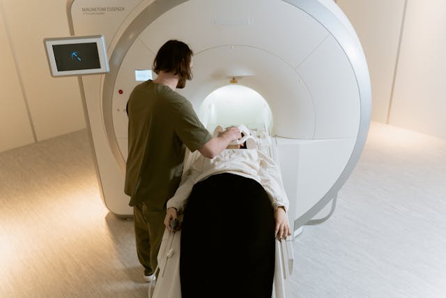
The Cambridge team examined data from 585 adults aged 40 to 59 living across the UK and Ireland, who were participating in the PREVENT-Dementia programme, which follows people in midlife to identify early risk factors. Participants were assessed on several measures: the level of neighbourhood deprivation based on their postcode; their performance on cognitive tests; lifestyle risk factors such as sleep quality, physical activity, weight, and blood pressure; and MRI scans to detect any early signs of damage in the brain’s small vessels. By comparing these factors, the researchers were able to explore how environmental disadvantage may influence brain health both directly and indirectly.

The study showed that individuals living in areas with higher unemployment rates, lower incomes, or fewer educational opportunities were more likely to experience difficulties maintaining healthy lifestyle habits. They were more likely to have poor sleep, higher blood pressure, obesity, and lower physical activity levels. All of these are known contributors to dementia risk because they can damage the brain’s vascular system over time. Interestingly, people in more deprived communities tended to drink less alcohol than those in more affluent neighbourhoods, reminding us that health patterns vary across contexts and that public health strategies must be tailored accordingly.

Neighbourhood deprivation was also associated with reduced cognitive performance, particularly in processing speed, spatial awareness, and concentration. These skills are vital for everyday functioning and often decline early in the path toward dementia. The researchers believe this reduced cognitive ability may be tied to the vascular damage seen on brain scans. When small vessels are weakened or obstructed, the brain receives less oxygen and fewer nutrients, which can gradually impair neural function. Crucially, the findings showed that these effects were not simply a matter of individual education levels or personal background. Even people with university degrees and stable employment were affected when they lived in deprived areas, highlighting how the physical and social environment can shape behaviour and health in powerful ways.

The results of this study emphasise that dementia prevention must go beyond encouraging individuals to make healthier lifestyle choices. While personal habits are essential, people can only act within the possibilities their environment allows. If someone lives in a neighbourhood without safe public spaces, access to affordable, healthy food, reliable healthcare, or a sense of security, maintaining good health becomes far more difficult. The researchers argue that meaningful dementia prevention will require policymakers to address the social conditions that limit healthy living. Improving neighbourhood safety, creating accessible exercise spaces, reducing economic inequality, and strengthening community support systems could all help lower dementia risk at a population level. In short, the study reinforces the idea that where we live deeply matters for how our brains age.

More information: Audrey Low et al, Neighborhood deprivation and midlife cognition: Evidence of a modifiable vascular pathway involving health behaviors and cerebral small vessel disease, Alzheimer’s & Dementia. DOI: 10.1002/alz.70756

Journal information: Alzheimer’s & Dementia Provided by University of Cambridge

Fresh study reveals: neurons build ‘air-tunnel’ networks that circulate harmful proteins linked to Alzheimer’s disease

Scientists at Johns Hopkins Medicine have discovered that mammalian brain cells can form microscopic tubes that transport toxic molecules between neurons, functioning like tiny “pneumatic tubes” within the brain. This process, observed in experiments using genetically engineered mice, helps explain how harmful proteins linked to Alzheimer’s disease might spread from one cell to another. The study, funded by the National Institutes of Health and published on 2 October in Science, could change how researchers think about the brain’s internal communication systems and open new directions for treating neurodegenerative disorders.

In their experiments, the researchers found that neurons form slender channels called nanotubes to remove small toxic molecules. One of them is amyloid-beta, a sticky protein that forms plaques that mark Alzheimer’s disease. Hyungbae Kwon, an associate professor of neuroscience at Johns Hopkins, explained that while these tubes help cells expel toxins, they also facilitate their spread. “Cells have to get rid of toxic molecules,” Kwon said, “but by producing a nanotube, they can transmit this toxic molecule to a neighbouring cell. Unfortunately, this also spreads harmful proteins through the brain.”

Using high-powered microscopes and live-cell imaging, the team watched neurons extend long, finger-like projections that connected one cell’s dendrites — the branch-like arms — to another’s. These structures, named dendritic nanotubes, acted as highways for transporting small molecules, calcium, and even toxic proteins. The researchers noted that the shape and flexibility of the tubes enabled them to rapidly transfer information and materials between cells that were not directly touching, revealing a form of communication previously hidden from scientists.

To explore this process further, the team built computer models to simulate how nanotubes contribute to the buildup of amyloid-beta early in Alzheimer’s disease. Their findings showed that the brain may have an extra “connectivity layer” of nanotubes that works alongside synapses — the traditional communication points between neurons. This suggests that the brain’s internal network is more physically linked than previously thought, and that these nanotubes might play a role in both maintaining and disrupting brain health.

The researchers examined brain tissue from healthy mice and from mice genetically modified to develop Alzheimer ‘s-like plaques. They discovered that diseased mice had more nanotubes at 3 months of age, before symptoms appeared, than healthy mice. By six months, the difference between the two groups had lessened. This pattern suggests that nanotube formation increases during the early stages of disease, possibly facilitating the rapid transport of toxic proteins through brain tissue. When the scientists studied human brain samples using publicly available electron microscopy data, they found nanotubes forming between neurons, a pattern similar to that observed in mice, suggesting that this mechanism exists in both humans and mice.

Kwon and his team now plan to test whether other brain cells, such as glial cells, also form nanotubes. They hope to learn whether controlling the creation of these tubes could help prevent or slow diseases like Alzheimer’s. If researchers can find a way to adjust nanotube formation — increasing it to remove waste or reducing it to stop the spread of toxins — they might one day use this discovery to protect brain cells from degeneration. This new understanding of how the brain moves molecules around adds an intriguing piece to the puzzle of how neurodegenerative diseases begin and progress. It offers hope for more targeted, effective treatments in the future.

More information: Hyungbae Kwonet al, Intercellular communication in the brain through a dendritic nanotubular network, Science. DOI: 10.1126/science.adr7403

Journal information: Science Provided by Johns Hopkins Medicine

Guiding Screens and Streams: Grandparents’ Role in Children’s Media Engagement

A new study from Rutgers University has found that grandparents play a far more important role in managing children’s media use than previously recognised. Conducted by Dafna Lemish, a Distinguished Professor of Journalism and Media Studies at Rutgers, along with Galit Nimrod and Nelly Elias from Ben-Gurion University of the Negev, the research explores how grandparents act as mediators in their grandchildren’s digital lives. Published in the Journal of Aging Studies, the study introduces the idea of “secondary mediation,” a process where grandparents use strategies and rules created by parents to guide children’s media habits. The researchers argue that traditional mediation theories should be expanded to include this shared, intergenerational approach to caregiving.

The study highlights that grandparents often follow parental guidelines when supervising television, online content, and games, effectively reinforcing family values around media use. However, this cooperation depends significantly on the relationship between the parents and grandparents, as well as the grandparents’ own familiarity with technology. The authors found that this collaboration strengthens intergenerational bonds, turning media supervision into a shared effort rather than a source of conflict. It also demonstrates that grandparents are not just passive observers in childcare but active partners who help maintain consistency in children’s digital routines.

Professor Lemish explained that much of the research on children’s media consumption has focused mainly on parents, overlooking grandparents who are frequently involved in caregiving. She wanted to know whether grandparents used the same strategies as parents—restricting screen time, discussing content, and co-viewing—or were more relaxed, perhaps “spoiling” their grandchildren. As both a scholar and a grandmother, Lemish was personally drawn to understanding these dynamics. Her findings reveal that grandparents do engage in media mediation, though often in more informal and emotionally supportive ways, reflecting their unique role in family life.

The research found that grandparents can have a positive influence by reducing children’s exposure to harmful content and encouraging media that supports learning and creativity. Lemish noted that grandparents can help counteract adverse effects such as exposure to violence, bullying, or stereotypes, while promoting enjoyment and intellectual growth. This balance of caution and encouragement allows children to benefit from the educational and social advantages of media without overindulgence. Grandparents’ wisdom and experience, the study suggests, can help young children navigate digital spaces more safely and thoughtfully.

A key recommendation from the researchers is that parents and grandparents should communicate openly about media rules and expectations. When caregivers share similar values and guidelines, children are less likely to experience confusion or tension. Lemish advised families to discuss their perspectives on media, agree on limits for screen time, and decide together when exceptions might be acceptable. This cooperative approach ensures that children receive consistent messages about using media responsibly while still allowing for flexibility and enjoyment.

To reach their conclusions, the researchers surveyed 267 pairs of grandmothers and mothers of children aged four to eight. They discovered that grandmothers were more confident in mediating noninteractive media, such as films or television, than interactive media, such as games. Factors such as education, time spent caregiving, and comfort with technology influenced how they guided their grandchildren. Overall, the study provides some of the first detailed insights into how grandparents shape children’s media habits. It portrays them as vital partners in helping families find balance in an age where screens are a constant presence in everyday life.

More information: Galit Nimrod et al, Secondary mediation of children’s media use: An exploration of mothers-grandmothers’ mediation dynamic, Journal of Aging Studies. DOI: 10.1016/j.jaging.2025.101357

Journal information: Journal of Aging Studies Provided by Rutgers University

Alterations in brain structure may reveal early indicators of dementia

A new study from the University of California, Irvine’s Center for the Neurobiology of Learning and Memory has found that ageing changes the brain’s shape in ways that can be measured and linked to mental decline. Rather than focusing only on the size of individual regions, researchers used a new analytic method to track how the brain’s overall form shifts and distorts over time. Their findings reveal that these geometric changes could provide early warning signs of cognitive impairment.

The research, published in Nature Communications and supported by the National Institute on Aging, showed that the brain’s shape changes systematically with age. The study’s senior author, Dr Niels Janssen of Universidad de La Laguna and UC Irvine, explained that while most studies focus on tissue loss, this one uncovered how the brain’s entire structure moves and warps in patterns tied to memory and reasoning decline. These shifts, he noted, could be a powerful new way to gauge brain health.

Using more than 2,600 brain scans from adults aged 30 to 97, the researchers found that some areas of the brain expand outward while others shrink inward with age. Specifically, the inferior and anterior areas of the brain tended to bulge, whereas the superior and posterior regions compressed. This uneven reshaping was most pronounced in people showing signs of cognitive decline, and those with greater posterior compression often performed worse on reasoning tasks. The same pattern appeared across multiple datasets, confirming the reliability of these changes as a hallmark of ageing.

One particularly intriguing finding involves the entorhinal cortex, a small but vital memory centre in the brain. The study suggests that as the brain reshapes over time, this region may be physically pushed closer to the hard base of the skull. This compression could make it more vulnerable to damage, particularly from tau, the toxic protein associated with Alzheimer’s disease. The research team proposes that these mechanical stresses may partly explain why this region is often one of the first affected in Alzheimer’s – a new way of thinking about how the disease begins.

Dr Michael Yassa, co-author and director of UC Irvine’s centre, said this discovery could change how scientists view early Alzheimer’s detection. He explained that if brain shape changes gradually increase pressure on fragile regions, it could trigger the cascade of damage that leads to dementia. Identifying these geometric markers early could one day help detect risk years before symptoms appear, providing a valuable window for intervention.

The study, a collaboration between UC Irvine and Universidad de La Laguna, highlights the promise of using brain geometry as a new diagnostic tool. As Dr Janssen noted, this research goes beyond simply measuring brain shrinkage—it shows how the brain’s architecture evolves with age and predicts who may be at higher risk for cognitive decline. By uncovering these patterns, scientists may be closer to understanding the physical forces that shape the ageing brain and its connection to diseases like Alzheimer’s.

More information: Yuritza Y. Escalante et al, Age-related constraints on the spatialgeometry of the brain, Nature Communications. DOI: 0.1038/s41467-025-63628-3

Journal information: Nature Communications Provided by University of California – Irvine

Gene Responsible for Familial Vision Loss Found

A team of scientists from the Medical University of Vienna and the Medical University of Graz has uncovered a new genetic cause of hereditary optic atrophy. This degenerative disease leads to gradual vision loss. Their findings, recently published in Genetics in Medicine, offer new opportunities for diagnosis and research into the underlying biological processes responsible for the condition. This discovery represents a significant step forward in understanding why some families are affected by inherited vision loss.

The research began with the study of a large Austrian family in which seven members across three generations suffered from optic atrophy. Genome-wide sequencing revealed a previously unknown mutation in the PPIB gene, which encodes an enzyme that helps proteins fold correctly and removes defective ones. In cells from affected individuals, this mutation was found to disrupt mitochondrial function—the “power plants” of cells that provide the energy needed for healthy vision.

Further analysis of existing genetic databases led the team to identify 12 additional individuals from 8 unrelated families who shared the same PPIB mutation, confirming its link to the disease. According to study leader Professor Wolfgang M. Schmidt of MedUni Vienna, the discovery establishes PPIB as a new optic atrophy gene. His colleague, Dr Thomas P. Georgi of MedUni Graz, highlighted that this finding allows for more precise genetic diagnoses and better support for affected families, helping clinicians offer more accurate advice and care.

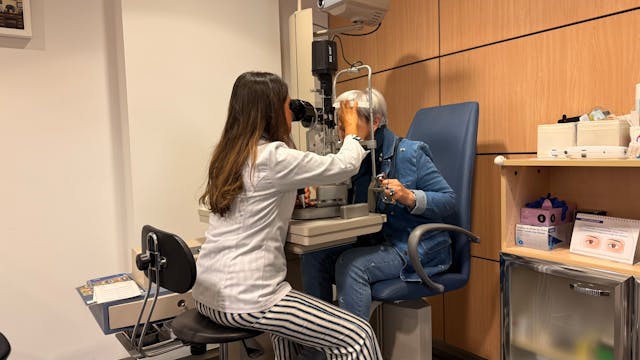
Optic atrophy damages the optic nerve, which transmits signals from the retina to the brain. It causes a gradual loss of visual clarity, reduced colour perception, and central blind spots. Although around twenty genetic forms of optic atrophy are currently known—most related to mitochondrial dysfunction—about 60 per cent of patients still lack a confirmed genetic explanation. Identifying PPIB helps fill this gap, giving researchers a new clue to explore and patients a more accurate understanding of their condition.

Beyond its relevance to optic atrophy, the discovery of the PPIB mutation could shed light on other diseases linked to mitochondrial damage and protein misfolding. As genome sequencing becomes increasingly available, testing for PPIB variants may soon become part of standard genetic screenings for unexplained vision loss. This breakthrough, therefore, not only advances scientific knowledge but also holds real promise for improving diagnosis, care, and future treatment of inherited eye diseases.

More information: Thomas P. Georgi et al, A recurrent missense variant in the PPIB gene encoding peptidylprolyl isomerase B underlies adult-onset autosomal
dominant optic atrophy, Genetics in Medicine. DOI: 10.1016/j.gim.2025.101595

Journal information: Genetics in Medicine Provided by Medical University of Vienna

New guidelines reveal a sharp rise in the number of American adults classified as obese

A new definition of obesity released earlier this year by the Lancet Diabetes and Endocrinology Commission could dramatically increase the number of Americans classified as obese. Researchers at Mass General Brigham found that under the updated criteria—which include measures of body fat distribution alongside the traditional body mass index (BMI)—the prevalence of obesity jumped from about 40 per cent to nearly 70 per cent in a study of more than 300,000 adults. The increase was especially notable among older individuals, and those newly classified as obese also faced higher risks of poor health outcomes. The findings were published in JAMA Network Open.

Dr Lindsay Fourman, co-first author and endocrinologist at Mass General Brigham, described the results as “astounding,” noting that the United States may now have an even greater obesity epidemic than previously understood. “With potentially 70 percent of the adult population now considered to have excess fat, we need to better understand what treatment approaches to prioritise,” she said.

Traditionally, obesity has been assessed using BMI, a simple ratio of weight to height. However, this measure fails to distinguish between fat and muscle mass or to account for where fat is stored in the body. The new framework expands on BMI by incorporating other anthropometric measures, such as waist circumference, waist-to-height ratio, and waist-to-hip ratio, which can better reflect unhealthy fat distribution.

According to the updated definition, a person is considered obese if they have a high BMI plus one elevated measure of body fat (“BMI-plus-anthropometric obesity”), or if they have a normal BMI but at least two elevated fat distribution measures (“anthropometric-only obesity”). The definition also distinguishes “preclinical” obesity from “clinical” obesity, the latter involving physical impairments or organ dysfunction associated with excess fat. Over seventy organisations, including the American Heart Association and The Obesity Society, have endorsed this new framework.

The researchers analysed data from over 300,000 participants in the National Institutes of Health’s All of Us Research Program. Under the new definition, obesity prevalence rose to 68.6 per cent, compared to 42.9 per cent using BMI alone. The change was primarily due to individuals classified as having anthropometric-only obesity, with obesity rates reaching nearly 80 per cent among adults over 70. Crucially, these newly identified individuals also faced higher risks of diabetes, cardiovascular disease, and early death compared to people without obesity.

Senior author Dr Steven Grinspoon emphasised that BMI has long been a limited tool for understanding body composition. The discovery that people with normal BMI but high body fat have elevated health risks raises new questions about treatments and preventive care. The team plans further research into therapies targeting fat distribution, including one that reduces waist circumference. As Dr Fourman explained, “Body composition matters—it’s not just pounds on a scale.”

More information: Lindsay Fourman et al, Implications of a New Obesity Definition Among the All of Us Cohort, JAMA Network Open. DOI: 10.1001/jamanetworkopen.2025.37619

Journal information: JAMA Network Open Provided by Mass General Brigham