Monthly Archives: December 2024

Scorbutus: Not Merely a Disease of 18th-Century Mariners

Scurvy, also known as vitamin C deficiency, is not solely a historical ailment confined to 18th-century sailors, as illustrated by a case involving a 65-year-old woman with limited mobility and social isolation. This case is detailed in an article published in the Canadian Medical Association Journal (CMAJ), found at CMAJ. Medical professionals highlight the importance of considering scurvy as a possible diagnosis in patients who exhibit abnormal bleeding and nonspecific symptoms.

The individual in question sought medical help at an emergency department in a downtown Toronto hospital, presenting with symptoms such as leg pain and weakness, skin lesions, and discolouration. Her medical history included several chronic health conditions, and her physical disabilities severely limited her ability to shop for groceries, cook, and carry out other daily tasks. With minimal external support, her diet primarily consisted of canned soup and fish, devoid of fresh fruits or vegetables.

Dr. Sarah Engelhart, a general internist at Mount Sinai Hospital and the University of Toronto, expressed that this case exemplifies the complex nature of food insecurity leading to rare diagnoses. A comprehensive evaluation of the patient’s social and dietary history was crucial in reaching a conclusive diagnosis.

Despite common perceptions, vitamin C deficiency remains surprisingly prevalent in the modern era. In the United States, about 5.9% of the population is affected, and in certain socioeconomically disadvantaged groups in the United Kingdom, the rate may reach as high as 25%.

The challenge in diagnosing vitamin C deficiency is its vague symptoms, including fatigue, weakness, and shortness of breath. The patient’s smoking habit further exacerbated her deficiency. Following the initiation of vitamin C therapy, her symptoms improved significantly, and a subsequent blood test confirmed the deficiency.

Medical professionals are advised to remain vigilant for signs of vitamin C deficiency when evaluating patients, particularly among vulnerable groups such as children, elderly individuals living in isolation, those with restrictive eating habits like those seen in autism spectrum disorder or individuals subsisting on a ‘tea and toast’ diet, smokers, substance users, or those suffering from malabsorption syndromes. The study’s authors stress the importance of being aware of food insecurity, a significant risk factor that impacts approximately one in five Canadian households.

More information: Kevin R. Murray et al, Scurvy in a 65-year-old woman with severely limited function and social supports, Canadian Medical Association Journal. DOI: 10.1503/cmaj.240769

Journal information: Canadian Medical Association Journal

Deciphering Time: Circadian Rhythms and Their Impact on Inflammatory Bowel Diseases

This study highlights the critical role of the body’s circadian clock in modulating immune responses and digestive functions, illustrating its significant influence on the onset and progression of inflammatory bowel diseases (IBDs) such as Crohn’s disease and ulcerative colitis. The findings indicate that disturbances in circadian rhythms can exacerbate inflammation, suggesting that synchronising treatment approaches with these inherent biological cycles could improve patient outcomes. The research advocates for a personalised approach to medicine that incorporates individual chronotypes alongside lifestyle interventions such as sleep hygiene and meal timing and employs real-time biological monitoring. This approach promises to revolutionise therapy for IBD, tackling both the physical and emotional burdens of these chronic conditions.

Insights from a review by Prof. Oren Froy of Hebrew University and Prof. Yael Weintraub from Schneider Children’s Medical Center and Tel Aviv University shed light on the crucial link between the body’s circadian clock and IBD. This connection opens up new avenues for innovative therapeutic strategies. The study demonstrates that both immune system activity and digestive functions are regulated by daily rhythms driven by the circadian clock. The disruption of this internal timing system is linked to increased inflammation, underscoring its pivotal role in the exacerbation and development of IBD.

Despite advancements in understanding the molecular mechanisms controlling the circadian clock, translating this knowledge into practical clinical applications remains challenging. Prof. Froy highlights the complexity of applying theoretical insights to practical treatment solutions, indicating the need for further research to bridge this gap.

Inflammatory bowel diseases, including Crohn’s disease and ulcerative colitis, impact approximately 10 million people worldwide, with their prevalence steadily increasing across both developed and developing countries. These conditions severely disrupt patients’ lives by causing symptoms such as abdominal pain, persistent diarrhoea, fatigue, and malnutrition, which significantly impair their quality of life. Beyond the physical symptoms, IBDs impose substantial emotional and financial burdens on sufferers, often leading to anxiety, depression, and limitations in work or social activities.

The study authors advocate for well-designed studies to deepen our understanding of how the circadian clock influences inflammation and IBD. Such research is crucial for developing personalised treatment strategies that consider individual chronotypes—natural preferences for activity and sleep patterns—and utilise wearable devices for monitoring circadian rhythms.
Looking ahead, the study outlines several key points and future directions for IBD management: personalised medicine that tailors therapies to individual patient needs, biological sampling and monitoring to gain insights into circadian impacts on inflammation, lifestyle interventions to reset the circadian clock, and the development of tools to evaluate circadian system performance. These strategies could enhance current treatments’ effectiveness and offer a new paradigm in IBD therapy.

Dr. Weintraub concludes, “This research opens doors to integrating circadian biology into IBD management, offering hope for therapies that not only address symptoms but also align with the body’s natural rhythms.” The promise of these rhythm-based therapies could significantly change the landscape of IBD treatment, providing relief and improved quality of life for millions affected by these challenging conditions.

More information: Yael Weintraub et al, The circadian clock in inflammatory bowel diseases, Trends in Molecular Medicine. DOI: 10.1016/j.molmed.2024.11.008

Journal information: Trends in Molecular Medicine Provided by The Hebrew University of Jerusalem

Assessing Unhealthy Alcohol Consumption Before It’s Too Late

A recent study suggests that a blood test could be a more effective method of identifying liver disease compared to traditional inquiries about a person’s alcohol consumption.
The common practice among clinicians when screening for potential alcohol-related liver damage involves simply questioning patients about their drinking habits. Despite its frequency of use, this approach might not be the most reliable. Researchers are now pointing towards a blood test as a superior alternative for detecting whether alcohol consumption is affecting the liver, potentially allowing for timely medical intervention to prevent severe damage.

At the University of California, San Francisco, experts have identified a biomarker known as phosphatidylethanol (PEth), which could offer a more accurate assessment of a person’s alcohol intake and its impact. This biomarker helps in understanding the risk of liver fibrosis, a condition characterized by the accumulation of scar tissue in the liver, which can progress to cirrhosis, liver failure, and even liver cancer if left untreated.

“The direct measurement of alcohol’s impact through PEth is a clearer and more direct method than the subjective accounts from patients about their drinking,” explains Judy Hahn, PhD, a professor in the Division of HIV, Infectious Diseases and Global Medicine at UCSF. She likens this approach to how clinicians handle other health indicators: “We don’t simply ask someone how much fatty food they eat – we measure their cholesterol. Similarly, we don’t ask people to estimate their weight – we measure it.”

The study, published in the American Journal of Gastroenterology, compared PEth levels with another liver disease risk indicator, the Fibrosis 4 (FIB-4) score. FIB-4 is calculated using a person’s age and results from several other blood tests. The findings indicate that while PEth levels closely correlate with FIB-4, the association between FIB-4 and self-reported alcohol consumption was considerably weaker. This discrepancy could be attributed to participants underreporting or forgetting the quantity of alcohol they consume.

This extensive study, which included over 4,000 participants from the United States, Russia, Uganda, and South Africa, is the largest to date to examine the relationship between PEth levels and liver fibrosis risk. It is also the first to assess the effectiveness of PEth compared to self-reported alcohol use in predicting fibrosis risk.

The progression of liver fibrosis can be slowed or even reversed with lifestyle changes, mainly through reducing alcohol consumption and improving diet. Early detection is vital to managing the condition effectively before it develops into more advanced stages of liver disease.

The researchers propose that PEth screening could become part of routine blood tests that check for cholesterol and blood sugar levels. “To effectively prevent and manage liver fibrosis, it’s crucial to accurately understand how much alcohol a person consumes,” states Pamela Murnane, PhD, MPH, an assistant professor of epidemiology and biostatistics and the lead author of the study. “Self-reports are proving insufficient for this purpose.” This advancement in medical screening could revolutionize how clinicians approach the diagnosis and treatment of liver disease related to alcohol consumption.

More information: Murnane, Pamela M. et al, Using Phosphatidylethanol as an Adjunct to Self-Reported Alcohol Use Improves Identification of Liver Fibrosis Risk, The American Journal of Gastroenterology. DOI: 10.14309/ajg.0000000000003178

Journal information: The American Journal of Gastroenterology Provided by University of California – San Francisco

Social Inequality Tied to Declining Brain Health in the Elderly and Dementia Cases

Researchers at Trinity College Dublin have engaged in a groundbreaking collaborative effort with global partners to investigate whether societal inequality impacts brain health. Their findings are detailed in a research paper published in Nature Aging today, Friday, December 27th. The study was conducted by an international team from the Multipartner Consortium to expand dementia research in Latin America (ReDLat), the Latin American Brain Health Institute (BrainLat), the Global Brain Health Institute (GBHI) at Trinity College Dublin, and other centres worldwide. It establishes a clear correlation between structural inequalities, such as socioeconomic disparities measured by the country-level GINI index, and alterations in brain structure and connectivity typical of ageing and dementia.

The research particularly highlights how societal inequities are biologically embedded, mainly affecting underrepresented groups in Latin America and the United States. The study presents several critical findings:

Firstly, the researchers observed that greater levels of inequality correlate with diminished brain volume and disrupted connectivity, particularly affecting the temporal-posterior and cerebellar regions crucial for memory and cognitive functions. These impacts were notably more severe in Latin America, underscoring the particular susceptibility of these populations to broad socioeconomic stressors.

Secondly, the study disclosed that Latinos suffering from Alzheimer’s disease endure the most drastic effects. This suggests that the environmental pressures associated with structural inequality might intensify neurodegeneration in ageing populations. In contrast, the milder impacts noticed in cases of frontotemporal lobar degeneration suggest a more substantial genetic influence in this condition. It is important to note that reduced brain volume and connectivity, standard in dementia patients, are linked with the progression and severity of the disease.

Furthermore, the research established that these associations persisted even after accounting for individual factors such as education, age, sex, and cognitive ability. This underscores the independent influence of macro-level factors on brain health, demonstrating that living in an environment of widespread inequality impacts brain health irrespective of one’s specific socioeconomic status. Thus, the research reveals the extensive effects of societal disparities on brain function.

Dr Agustina Legaz, PhD from the ReDLat consortium and the study’s first author, emphasised the critical need to incorporate both individual social determinants of health and broader exposome factors, like social and physical variables, into global brain health research. She highlighted that these findings pave the way for future studies exploring the biological mechanisms linking aggregate inequality to ageing and neurodegeneration.

Dr Agustín Ibáñez, a professor in global brain health at Trinity College and director of BrainLat, also the corresponding author, remarked on the significant role of structural inequality in shaping brain health. He pointed out that with dementia rates rising, particularly in low- and middle-income countries, the study’s findings stress the need for targeted interventions to address the root causes of brain health disparities, which are specific to each region.

The study advocates for a multi-level approach to achieving brain health equity, which involves exploring the biological embedding of other macro-level exposome factors beyond socioeconomic inequality. These could include democratic governance, air pollution, migration, climate change, and access to green spaces. By identifying and addressing these region-specific modulators, targeted interventions could be developed to slow down brain ageing and reduce the burden of dementia in disadvantaged communities.

More information: Agustina Legaz et al, Structural inequality linked to brain volume and network dynamics in aging and dementia across the Americas, Nature Aging. DOI: 10.1038/s43587-024-00781-2

Journal information: Nature Aging Provided by Trinity College Dublin

Research Reveals How Elevated Blood Sugar Heightens Thrombosis Risk

A study conducted at the Center for Research on Redox Processes in Biomedicine (Redoxoma) sheds light on how high blood sugar (hyperglycemia), a key symptom of diabetes, can contribute to thrombosis. Published in the Journal of Thrombosis and Haemostasis, the findings offer valuable insights that could inform the development of preventive strategies for cardiovascular diseases in individuals with diabetes.

According to Renato Simões Gaspar, the first author of the study, cardiovascular events like heart attacks and strokes, which are common causes of death in Brazil and other Latin American countries, are often precipitated by arterial thrombosis. Hyperglycemia, along with other risk factors such as dyslipidemia and hypertension, is mainly associated with an increased risk of cardiovascular diseases. “Hyperglycemia appears to be significantly linked to cardiovascular dysfunction,” said Gaspar.

The research was supported by FAPESP and carried out during Gaspar’s postdoctoral research under the leadership of Francisco Laurindo, the last author of the article. Laurindo, a professor at the University of São Paulo’s Medical School (FM-USP) and a member of Redoxoma, collaborated with Gaspar in this investigation. Redoxoma is a Research, Innovation, and Dissemination Center (RIDC) established by FAPESP at the Institute of Chemistry (IQ-USP). Gaspar is a faculty member at the State University of Campinas (UNICAMP).

The study focuses on the relationship between prolonged hyperglycemia, diabetic ketoacidosis, and an increased risk of thrombosis. These conditions cause endothelial dysfunction, disrupting the blood vessel lining, which plays a pivotal role in platelet adhesion and thrombus formation. This mechanism underpins the heightened susceptibility to thrombosis seen in individuals with diabetes.

The research specifically explored the role of peri/epicellular protein disulfide isomerase A1 (pecPDI) in regulating platelet-endothelium interactions in hyperglycemia. The study revealed that pecPDI, through adhesion-related proteins and changes to endothelial membrane biophysics, is critical in mediating thrombosis in diabetic conditions. “We discovered that a pathway for PDI in endothelial cells mediates thrombosis in diabetes when hyperglycemia is present, involving a specific molecular mechanism that we identified,” said Laurindo.

PDI, an enzyme located in the endoplasmic reticulum, facilitates the formation of disulfide bridges in nascent proteins, ensuring that they fold into their correct three-dimensional structure. PecPDI, a form of PDI found in the extracellular space, is secreted or bound to the cell surface in various cells, including platelets and endothelial cells. Previous studies have shown that pecPDI regulates thrombosis across different models.

To delve deeper into the interaction between platelets and endothelial cells under hyperglycemic conditions, the researchers developed a model using human umbilical vein endothelial cells cultured under different glucose concentrations. This model generated both normoglycemic and hyperglycemic cells, allowing the team to assess the impact of PDI on platelet adhesion using PDI or pecPDI inhibitors. The results were striking: platelets adhered almost three times more to hyperglycemic cells than to normoglycemic ones. However, inhibiting PDI reversed this effect, suggesting that pecPDI plays a critical role in platelet-endothelium interactions during hyperglycemia.

To further understand these findings, the researchers examined biophysical changes such as cytoskeleton remodelling in endothelial cells. They discovered that hyperglycemic cells exhibited more structured actin filament fibres than normoglycemic cells. Additionally, they measured hydrogen peroxide production, as reactive oxygen species are key mediators of cytoskeleton reorganisation and cell adhesion—hyperglycemic cells produced twice as much hydrogen peroxide as their normoglycemic counterparts.

The team also explored whether cytoskeleton reorganisation affected cell membrane stiffness, influencing platelet adhesion. Using atomic force microscopy, they confirmed that hyperglycemic cells were stiffer than normoglycemic ones. The microscope images revealed elongations in the cells, accompanied by extracellular vesicles that appeared to detach from the elongations. This prompted the researchers to investigate the secretome—the collection of proteins secreted by cells into the extracellular space—to determine if these vesicles contained proteins that promoted platelet adhesion.

The researchers identified 947 proteins in the secretome of hyperglycemic cells, of which eight were involved in cellular adhesion. Using RNA interference to silence gene expression for three of these proteins, they found that two proteins, SLC3A2 and LAMC1, played key roles in modulating platelet adhesion. SLC3A2 is a membrane protein, while LAMC1 is a subunit of laminin 1, a critical extracellular matrix component.

The study’s overall conclusion is that hyperglycemia triggers the secretion of specific proteins that promote platelet adhesion. Inhibition of PDI and pecPDI prevented endothelial cells from secreting these proteins, offering a potential target for therapeutic intervention to reduce thrombosis risk in diabetic patients.

This research underscores the complex interplay between hyperglycemia, endothelial dysfunction, and thrombosis. Identifying the molecular mechanisms involved opens up new avenues for preventing cardiovascular complications in individuals with diabetes, potentially leading to more effective treatments for reducing the burden of cardiovascular diseases in diabetic populations.

More information: Renato S. Gaspar et al, Endothelial protein disulfide isomerase A1 enhances membrane stiffness and platelet-endothelium interaction in hyperglycemia via SLC3A2 and LAMC1, Journal of Thrombosis and Haemostasis. DOI: 10.1016/j.jtha.2024.08.001

Journal information: Journal of Thrombosis and Haemostasis Provided by Fundação de Amparo à Pesquisa do Estado de São Paulo

Inconsistent Sleep Patterns Associated with Increased Risk of Serious Cardiovascular Incidents

An irregular sleep-wake cycle, even when fulfilling the recommended amount of sleep, is linked with an increased risk of major cardiovascular events, including heart attack and stroke. This association was revealed in a study published in the Journal of Epidemiology & Community Health, which delves deeper into the impact of sleep patterns on health, a relatively less explored area than sleep duration.

The research utilised data from 72,269 individuals aged between 40 and 79 who were participants in the UK Biobank study and had no prior history of significant cardiovascular events. These participants were monitored over a week with an activity tracker that recorded their sleep. This data helped calculate each individual’s Sleep Regularity Index (SRI) score. Based on the SRI scores, the participants were categorised into three groups: those with a score above 87 were deemed to have regular sleep patterns, scores below 72 indicated irregular sleepers and scores between these values were considered moderately irregular.

Over the following eight years, the study tracked incidents of cardiovascular mortality, heart attack, heart failure, and stroke using death registries and hospital records. This enabled researchers to correlate the risk of these cardiovascular events with the participants’ sleep patterns. After adjusting for several potential influencing factors such as age, lifestyle, diet, and mental health, the findings indicated that individuals with irregular sleeping patterns were 26% more likely to experience a major cardiovascular event compared to those with regular sleep patterns. Those with moderately irregular sleep patterns exhibited an 8% increased risk.

Further analysis highlighted a nearly linear relationship between lower SRI scores and higher risk of cardiovascular events, with a more significant decrease in risk noted at higher SRI scores. Interestingly, while 61% of regular sleepers met the recommended sleep duration of 7 to 9 hours for adults aged 18 to 64 and 7 to 8 hours for those over 65, only 48% of irregular sleepers achieved this. However, meeting the recommended sleep duration did not significantly mitigate the risk for irregular sleepers, though it did reduce the risk for moderately irregular sleepers.

It’s crucial to note that this observational study cannot definitively establish causality. The researchers acknowledged several limitations to their findings. For example, the sample size, although significant, may not perfectly reflect the broader UK population. Sleep patterns were assessed for one week, and the activity tracker could not differentiate between quiet wakefulness and actual sleep. Furthermore, considering only the most extended sleep period, the SRI algorithm did not account for napping.

Despite these limitations, the study’s results strongly suggest that irregular sleep significantly correlates with the risk of major adverse cardiovascular events in adults, regardless of meeting sleep duration recommendations. The findings underscore the importance of sleep regularity over mere duration in influencing cardiovascular health risks.
The researchers advocate for a greater focus on sleep regularity in public health guidelines and clinical practice, highlighting its potential role in cardiovascular health management. This shift in focus could lead to more effective strategies in preventing cardiovascular events, emphasising how long we sleep and how consistently we adhere to our sleep schedules.

More information: Jean-Philippe Chaput et al, Sleep regularity and major adverse cardiovascular events: a device-based prospective study in 72 269 UK adults, Journal of Epidemiology and Community Health. DOI: 10.1136/jech-2024-222795

Journal information: Journal of Epidemiology and Community Health Provided by BMJ Group

Assessing the Impact of Disease and Addressing Treatment Challenges in Primary Progressive Aphasia

Imagine the frustration of slowly losing the ability to express yourself, not due to a lapse in memory but because the words you wish to say stubbornly refuse to be spoken. This is the harsh reality faced by those afflicted with primary progressive aphasia (PPA), a relatively uncommon form of dementia that typically manifests in middle age and gradually deteriorates language abilities over time.

At the University of Chicago Medicine, a team of dedicated researchers is shedding light on the challenges encountered by individuals with PPA and are pioneering accessible treatment options. Their recent publications offer new insights into the substantial effect PPA has on quality of life and highlight the viability of telemedicine interventions on an international scale. This breakthrough might revolutionise care delivery and influence policy decisions.

PPA stands distinct in the spectrum of neurological disorders because it predominantly impairs language skills, unlike the more familiar Alzheimer’s dementia, which initially affects memory. Emily Rogalski, PhD, the Rosalind Franklin PhD Professor of Neurology at UChicago and a prominent figure in this research area, emphasises that PPA is often neglected in academic studies due to the difficulty in gathering large participant groups, which can obscure the true breadth of lived experiences.

Moreover, Rogalski points out the frequent oversight of PPA in clinical settings, particularly among individuals from lower socioeconomic backgrounds, leading to underdiagnosis and, consequently, a lack of appropriate care. This issue is compounded by the geographical and financial barriers many face when seeking specialised medical advice and treatment.

PPA’s early onset poses unique challenges. Rogalski notes, “Individuals with PPA are often still active in their careers and may be caring for young children at home.” This affects the patients and places a substantial burden on their families, affecting familial relationships and economic stability.

Rogalski and her team utilised the Health Utilities Index (HUI) to understand how PPA affects daily life. This well-established tool assesses various aspects of well-being, such as physical capabilities, emotional health, and cognitive functions. The findings confirmed the severe impact of PPA on health-related quality of life, correlating more significant language impairment with lower quality of life, particularly affecting hearing, sensation, cognition, and speech abilities.

Thomas Hopkins, PharmD, MS, the study’s lead author, explained that the research aimed not only to gather detailed data on the quality of life for those with PPA but also to enable direct comparisons with other diseases. This generic measurement tool is invaluable for policymakers and health resource allocators, ensuring comprehensive, comparative health metrics inform decisions.

This quality-of-life data is crucial for influencing decisions regarding research funding, insurance regulations, and disability coverage. It equips researchers, patients, and their families with the evidence needed to advocate for more substantial support and resources.

Concurrently, Rogalski and her colleagues have been exploring the potential of telemedicine in delivering speech-language therapy to PPA patients, a method that transcends geographic and economic barriers. Their study successfully enrolled 95 pairs of PPA patients and their primary caregivers from four countries, demonstrating the feasibility of using remote recruitment and video chat interventions.

This successful deployment of telemedicine facilitates equitable care and sets a potential model for future research and interventions across various dementia syndromes and conditions. Rogalski envisions this framework as adaptable for broader advocacy and intervention strategies.

Despite the lack of a cure for PPA, the role of care partners in these studies is pivotal. They provide essential perspectives on the everyday challenges and diverse needs that span family dynamics and various life situations, giving families a platform to voice their experiences.

Although challenges in diagnosing and treating PPA persist, and no curative therapy has been developed, these research advances offer hope and practical support to affected families. Rogalski passionately advocates for enhancing aspects of daily life that can be controlled, such as independence, emotional well-being, and confidence, while the search for pharmacological solutions continues. This approach provides hope and practical aid to those navigating the difficult journey of PPA, reinforcing the belief that improving quality of life in tangible ways is equally as crucial as pursuing a medical cure.

More information: Emily Rogalski et al, Communication Bridge-2 randomized controlled trial: Recruitment and baseline features, Alzheimer’s & Dementia. DOI: 10.1002/alz.14168

Journal information: Alzheimer’s & Dementia Provided by University of Chicago Medical Center

Scientists Discover that Disrupted Sleep Is a Key Symptom of the Most Prevalent Liver Disorder

Metabolic dysfunction-associated steatotic liver disease (MASLD), previously known as non-alcoholic fatty liver disease, is becoming increasingly common globally, exacerbated by rising obesity rates and sedentary lifestyles. MASLD is now the most prevalent liver disorder, affecting 30% of adults and 7% to 14% of children and adolescents. Projections suggest that by 2040, over 55% of adults may be affected. Individuals with MASLD are at a significantly increased risk of developing a range of serious health issues, including diabetes, hepatocellular carcinoma, other non-liver cancers, chronic kidney disease, sarcopenia (age-related muscle loss), and cardiovascular disease.

Previous research has pointed to disruptions in the circadian rhythm and sleep patterns as contributing factors in the onset of MASLD. The American Academy of Sleep Medicine, recognizing the need for more definitive evidence, has recommended the use of objective diagnostic methods over subjective tools like sleep questionnaires to establish the connection between sleep and circadian rhythm disorders and the development of MASLD and its more severe form, MASH (metabolic-associated steatohepatitis). MASH leads to liver inflammation and scarring due to excessive fat accumulation.

In a pioneering study, Dr Sofia Schaeffer, a postdoctoral researcher at the University of Basel and Basel’s University Center for Gastrointestinal and Liver Diseases, employed 24/7 actigraphy—a method that uses a wrist-worn sensor to track movement—to demonstrate distinct differences in the sleep-wake patterns of MASLD patients compared to healthy individuals. The findings, published in Frontiers in Network Physiology, revealed that MASLD patients experienced significantly more fragmented sleep due to frequent awakenings and increased wakefulness throughout the night.

From 2019 to 2021, Dr. Schaeffer and her team recruited 46 adults diagnosed with MASLD, MASH, or MASH with cirrhosis, 16 healthy volunteers, and eight patients with liver cirrhosis unrelated to MASH for comparison. Participants were outfitted with actigraphs that continuously monitored light exposure, physical activity, and body temperature. They were evaluated as outpatients at the start, midpoint, and end of a four-week follow-up period, during which they also maintained sleep diaries and completed sleep questionnaires.

The study found that while there were no significant differences in sleep duration or time spent in bed between MASLD patients and healthy individuals, MASLD patients woke up 55% more frequently and spent 113% more time awake after initially falling asleep. They also tended to sleep more during the day. Similar patterns were observed in patients with MASH and related conditions.

Subjectively, MASLD patients reported shorter, more disrupted sleep, often delayed in onset, with 32% noting sleep disturbances due to psychological stress—significantly higher than the 6% reported among healthy participants. Dr. Schaeffer concluded that sleep fragmentation appears to play a role in the pathogenesis of MASLD, though it remains unclear whether liver disease causes sleep disturbances or vice versa. The underlying mechanisms likely involve a combination of genetic, environmental, and immune response factors, all influenced by obesity and metabolic syndrome.

The study also included a single session on sleep hygiene to improve participants’ sleep patterns. Still, this intervention showed no significant impact on either actigraph-recorded or self-reported sleep quality and quantity. Dr Christine Bernsmeier, a professor at the University of Basel and the study’s senior author, suggests that future research should consider ongoing sleep counselling or interventions such as light therapy, combined with other lifestyle modifications, to enhance the sleep-wake cycles of individuals with MASLD. This comprehensive approach highlights the complex interplay between sleep, lifestyle factors, and liver health, underscoring the need for integrated treatment strategies to manage and mitigate the impacts of this increasingly prevalent liver disease.

More information: Sofia Schaeffer et al, Significant nocturnal wakefulness after sleep onset in metabolic dysfunction–associated steatotic liver disease, Frontiers in Network Physiology. DOI: 10.3389/fnetp.2024.1458665

Journal information: Frontiers in Network Physiology Provided by Frontiers

Solitude and Seclusion: Returning to Pre-Pandemic Figures Yet Remaining Elevated for Elderly Adults

Loneliness and isolation among older Americans have reverted mainly to pre-pandemic levels, indicating that over a third of individuals aged 50 to 80 still frequently experience these feelings, according to a recent national study. While the return to previous rates might initially seem optimistic, the fact remains that a significant portion of the elderly population continues to deal with feelings of loneliness and isolation. This issue is particularly pronounced among older adults facing substantial physical or mental health challenges, who report much higher levels of loneliness and isolation compared to their healthier counterparts.

The findings, which provide a deeper understanding of this issue, stem from six years of data collected by the National Poll on Healthy Aging. The research, led by the University of Michigan Institute for Healthcare Policy and Innovation and published in JAMA, highlights ongoing concerns. The poll, which began in 2018 and included the most recent data from earlier this year, surveys older adults across the United States about their experiences of loneliness—defined as the subjective feeling of being alone—and social isolation.

Recent data from 2024 show that 33% of older adults reported feeling lonely at least occasionally over the past year, mirroring the 34% reported in 2018. In contrast, loneliness peaked at 42% in the intervening years. Similarly, 29% of older adults reported feelings of social isolation in 2024, slightly higher than the 27% seen in 2018. The early stages of the COVID-19 pandemic saw this figure rise dramatically to 56%, though it has steadily decreased each year since.

Dr. Preeti Malani, the study’s lead author and a professor at the University of Michigan Medical School, notes that while returning to pre-pandemic levels may seem like progress, the baseline itself was problematic, particularly for specific groups of older adults who continue to suffer from very high rates of loneliness and social isolation. Dr Malani, who also served as a senior advisor to the poll, underscores the increased recognition of loneliness and isolation’s impact on health, especially as people age.

The study identifies groups with notably high rates of loneliness in 2024, including those who rate their mental health as fair or poor at 75%, an increase from 74% in 2018. Those rating their physical health as fair or poor also increased from 50% to 53%. Notably, those not working or receiving disability income (excluding retirees) saw a rise in loneliness from 38% to 52%. Similarly, high rates of social isolation were reported by the same groups, with those in poor mental health at 77%, up slightly from 79%, and those in poor physical health at 52%, up from 43%.

Jeffrey Kullgren, M.D., M.P.H., M.S., the poll’s director and an associate professor of internal medicine at the University of Michigan, emphasizes that clinicians should view loneliness and isolation as significant factors in the lives of their patients, especially those with serious health issues. Dr Kullgren advocates for screening for these conditions and connecting patients with community resources such as senior centres, Veterans’ groups, and volunteering opportunities. This approach addresses immediate social needs and contributes to the broader goal of enhancing the quality of life for older adults facing these challenges.

More information: Preeti N. Malani et al, Loneliness and Social Isolation Among US Older Adults, JAMA. DOI: 10.1001/jama.2024.23213

Journal information: JAMA Provided by Michigan Medicine – University of Michigan

Recent Study Suggests Daily Coffee Intake May Extend Healthy Lifespan by Up to Two Year

A newly published review paper, which appears in Ageing Research Reviews and has support from the Institute for Scientific Information on Coffee (ISIC), delves into the scientific connection between coffee and healthy ageing. The global demographic aged 65 and above is expanding rapidly, projected to increase from 10% in 2022 to 16% by 2050. This research emphasises how regular, moderate coffee consumption could play a crucial role for this segment of the population, contributing to a balanced and healthy lifestyle.

The European Food Safety Authority advises that up to 400mg of caffeine, equivalent to 3-5 cups of coffee daily, is a moderate and safe amount for adults. However, this intake should be halved to 200mg daily for pregnant or lactating women.

Coffee is one of the most extensively studied commodities globally, with over 50 studies identifying its potential to reduce all-cause mortality. This includes a discrete yet significant reduction in the risk of major health issues such as cardiovascular disease, cancer, respiratory diseases, cognitive decline, and frailty.

The review highlights that regular coffee drinking can extend a person’s healthy lifespan by an average of 1.8 years. This suggests a longer life and a healthier one. Interestingly, while some anti-ageing nutritional interventions may display a gender bias, the benefits of coffee consumption appear consistent across both men and women.

Further, the review explores coffee’s engagement in biological mechanisms associated with ageing, such as reducing genomic instability or cell mutations, which are key factors in ageing, and promoting normal cellular functions. It’s important to note that the research focused solely on human-based studies, ensuring a more accurate depiction of coffee’s impact on human health.

Historically, clinical guidelines for older adults have recommended reducing or avoiding coffee. However, this review calls for reconsidering these recommendations based on solid scientific evidence of coffee’s benefits in promoting healthy ageing.

Coffee’s health properties are due to its caffeine content and the presence of over 2,000 bioactive compounds. These include polyphenols, which offer antioxidant and anti-inflammatory benefits, potentially reducing neuroinflammation and improving insulin sensitivity.

While the beneficial effects of caffeine and its non-caffeine components in coffee are recognised, much remains to be understood about the precise mechanisms through which they influence health. The paper’s authors advocate for more comprehensive research into coffee’s health benefits.

Rodrigo Cunha, the lead author from the University of Coimbra, remarked on the significance of dietary interventions in a world where populations are ageing more rapidly than ever. He pointed out that traditional clinical advice often underestimates the role of coffee in healthy ageing. With substantial evidence suggesting that regular coffee consumption can mitigate the biological mechanisms that deteriorate with age, there is a clear need to revise existing dietary guidelines. Cunha stresses the importance of understanding how these interactions occur and identifying which individuals might benefit most from coffee’s effects.

More information: Cátia R. Lopes et al, Impact of coffee intake on human aging: Epidemiology and cellular mechanisms, Ageing Research Reviews. DOI: 10.1016/j.arr.2024.102581

Journal information: Ageing Research Reviews Provided by Kaizo

Homeownership in the US Associated with Increased Longevity

Dr Casey Breen, a Senior Postdoctoral Research Fellow at Oxford University’s Leverhulme Centre for Demographic Science and Department of Sociology, led a study recently published in Demography. This research investigated the correlation between homeownership and life expectancy in the United States, focusing on male Americans born in the early twentieth century. The findings indicated that homeownership contributed an additional 0.36 years to the life expectancy of Black males and 0.42 years for their White counterparts.

Dr. Breen remarked on the significance of these findings, noting, “My study demonstrates that homeownership significantly boosts life expectancy. These outcomes support the implementation of social policies aimed at broadening homeownership among Black Americans, which could potentially reduce the life expectancy disparities between Black and White males in the US.”

The research underscores the severe racial inequalities in mortality rates that persist in the US and points out the stark differences in homeownership rates between racial groups during the twentieth century. For example, during the 1940s, White Americans were nearly twice as likely to own a home as Black Americans. This disparity can be attributed to systemic historical challenges such as slavery and racial discrimination, which significantly hindered Black Americans’ ability to purchase homes. In 1940, fewer than 10% of Black Americans between 18 and 25 owned homes, and only 40% of those over 65 were homeowners.

To conduct this analysis, Dr. Breen utilised data from the 1920 and 1940 census records linked to social security mortality records. By employing a sibling-based identification strategy, the study effectively assessed the impact of homeownership on life expectancy among American males who owned homes between the ages of 24 and 35.

The research also explored other reasons homeownership might lead to longer life expectancies. Owning a home not only aids in wealth accumulation—often associated with better health outcomes—but also provides psychological benefits and fosters a sense of community, which are vital for well-being. Interestingly, the study noted that the property’s value had minimal influence on life expectancy, suggesting that the benefits of homeownership extend beyond financial aspects.

Furthermore, Dr. Breen highlighted another key finding: “There is a notable, statistically significant difference in life expectancy between Americans who own their homes and those who rent. Homeowners in early adulthood tend to live about six months longer at age 65 than those who rent.”

The study meticulously controlled factors such as educational attainment, race, income, marital status, and family background to understand better how homeownership impacted life expectancy in the US during the twentieth century. However, it is crucial to acknowledge that the study’s focus on specific demographics—gender, ethnicity, nationality, and historical context—means its findings may not be generalisable to other populations.

More information: Casey F. Breen et al, The Longevity Benefits of Homeownership: Evidence From Early Twentieth-Century U.S. Male Birth Cohorts, Demography. DOI: 10.1215/00703370-11680975

Journal information: Demography Provided by University of Oxford

Texas A&M Study Reveals Gut Healing Could Mitigate Long-Term Effects of Stroke

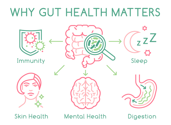
Researchers at Texas A&M University have uncovered that improving gut health could be crucial in enhancing long-term recovery for stroke patients. This finding was detailed in a recent paper published by the Department of Neuroscience and Experimental Therapeutics at the Texas A&M College of Medicine. The study represents a growing body of research that explores an innovative treatment approach, capitalising on the connection between the brain and digestive system to mitigate cognitive impairments and other prolonged consequences of stroke or brain trauma.

In their experiments, the research team discovered that a medication effective at protecting the brain immediately post-stroke did not alleviate long-term cognitive impairments when administered solely to the brain. However, when this drug was applied to the gut, it significantly reduced these impairments. This insight led to a surprising revelation for Regents Professor and Department Head Dr Farida Sohrabji, who noted, “Just fixing the brain directly won’t do it. As a neuroscientist, that was kind of shocking to me,” further emphasising the necessity of gut repair for improving long-term brain function.

The significance of these findings is detailed in the November edition of the journal Brain, Behavior, and Immunity. The paper builds upon prior research conducted under the leadership of Dr. Sohrabji, with contributions from graduate student Yumna El-Hakim and associate research scientist Dr. Kathiresh Kumar Mani. This team is particularly interested in how the brain and gut reciprocally influence each other during and after a stroke. By harnessing this relationship, they aim to develop new therapeutic methods to prevent cognitive decline in stroke survivors and lower their risk of progressing to dementia or Alzheimer’s disease (AD). Their research is supported by a grant from the National Institutes of Health and additional funding from the WoodNext Foundation.

Dr Sohrabji pointed out that stroke is a significant cause of dementia and AD, noting, “While there are acute, immediate consequences of stroke, there are also these long-term consequences that affect the quality of life for the patient as well as the caregivers, so there’s a lot of interest in understanding how to improve long-term outcomes.”

The dynamics within the gut following a stroke are critical yet underappreciated. Dr. Sohrabji described how, within moments of a stroke, there is evident immediate damage, such as paralysis of the arm or slurred speech. However, the significant damage to the intestine’s key structures is less noticeable at first, as the brain signals to the gut that something is awry. “We’ve found that minutes after a stroke occurs, normal gut anatomy is completely disrupted,” Dr. Sohrabji explained.

This disruption includes the breakdown of cells that keep the gut contents sealed from the rest of the body. This allows harmful digestive bacteria to leak out and potentially reach the brain or, at least, provoke a significant inflammatory response from the body. This inflammation can exacerbate the brain’s injury from the stroke, leading to increased long-term cognitive impairment.

The 2024 study highlighted the impact of treating the gut directly with Insulin-like Growth Factor (IGF-1), significantly reducing inflammation and cognitive impairment post-stroke. The treatment appeared to repair the damaged gut structures, supporting the idea that a healthy gut is vital for effective stroke recovery.

Additionally, the team is investigating the potential of stem cell transplants to rapidly repair the gut following a stroke, a method proposed by Dr Mani that has proven effective in earlier studies. Usually, the gut produces a steady supply of stem cells for self-repair. Previous research has shown that these cells can be transplanted from a healthy donor to a recipient with a damaged gut to expedite recovery.

Dr. Sohrabji shared their encouraging findings, “We were fairly sure that (the stem cells) would repair the gut. What was not known, and what was a very pleasant surprise to us, was that in that process, it also improved stroke outcomes.” She elaborated that the treatment reduced brain tissue death and preserved cognitive function following a stroke.

The team at Texas A&M continues to push forward with their research. They hope that their ongoing studies will lead to the development of a stem cell-derived treatment that could be administered to stroke patients soon after the event, thus reducing the long-term risks for dementia and other severe outcomes. Their innovative approach highlights the increasingly recognised importance of the gut-brain axis in medical science, particularly in the context of brain injuries and recovery.

More information: Yumna El-Hakim et al, Peripheral, but not central, IGF-1 treatment attenuates stroke-induced cognitive impairment in middle-aged female Sprague Dawley rats: The gut as a therapeutic target, Brain Behavior and Immunity. DOI: 10.1016/j.bbi.2024.08.008

Journal information: Brain Behavior and Immunity Provided by Texas A&M University