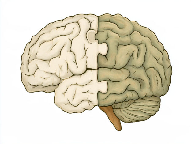
For centuries, scientists have viewed the brain as a single, unified organ. But new Stanford Medicine-led research suggests that the human brain develops from two distinct systems with separate origins, challenging a long-standing model of brain development.
Researchers had generally thought that a single progenitor cell early in development gives rise to the entire brain. The new findings suggest instead that the forebrain and midbrain develop from one type of progenitor cell, while the hindbrain follows a separate developmental pathway. These two systems appear to have evolved independently before eventually becoming joined together.
The distinction reflects the brain’s different functions. The forebrain supports higher-level abilities such as language, consciousness and abstract reasoning. The hindbrain, often associated with the brain stem, controls essential automatic functions including breathing, sleeping, heartbeat and hunger, as well as muscles involved in facial movement, speech and swallowing.
“We’ve shown for the first time that the front of the brain arises from a totally different progenitor cell than the back of the brain,” said Kyle Loh, PhD, associate professor of developmental biology and senior author of the study. The findings are being published in Nature Neuroscience, with graduate students Carolyn Dundes and Rayyan Jokhai as co-first authors.
The researchers made the discovery by studying mouse embryos during gastrulation, an early stage when the body begins to take shape. They identified two distinct populations of brain progenitor cells: cells expressing the gene Otx2 develop into the forebrain and midbrain, while those expressing Gbx2 form the hindbrain. The two populations remained separate from the earliest stages of development.
Further analysis revealed fundamental differences in how DNA is packaged within the two cell populations, effectively committing them to different developmental paths. This could explain why scientists have struggled for decades to produce human hindbrain neurons in the laboratory: previous approaches may have attempted to transform forebrain and midbrain progenitors into cells they were incapable of becoming.
Using their new understanding, the researchers successfully coaxed human pluripotent stem cells into functional hindbrain motor neurons in the laboratory. The cells displayed electrical activity and produced proteins characteristic of neurons controlling facial and swallowing muscles. This advance could provide new ways to study spinal muscular atrophy (SMA) and amyotrophic lateral sclerosis (ALS), conditions in which hindbrain neurons can progressively lose function.
The team also traced this two-part organisation through more than 550 million years of evolution, finding similar patterns in chickens, zebrafish and acorn worms. The discovery suggests that evolution may have brought two ancient neural systems together to form what we now recognise as the brain. The ability to grow hindbrain neurons in the laboratory could help researchers investigate brain-stem diseases, explore regenerative treatments and better understand other hindbrain functions, including the neural circuits involved in hunger.
More information: Rayyan Jokhai et al, Two parallel neural ectoderm progenitors contribute to the developing brain, Nature Neuroscience. DOI: 10.1038/s41593-026-02433-7
Journal information: Nature Neuroscience Provided by Stanford Medicine