Author Archives: support

Enhancing Longevity Through Improved Hand Dexterity

Among older adults, the decline in lower limb strength—necessary for walking, standing, and maintaining balance—has long been recognised as a primary contributor to the need for long-term or nursing care. Previous research by the same team established a strong link between reduced leg function and the likelihood of requiring assistance in later life. However, while the role of lower limbs in mobility has been extensively studied, far less attention has been paid to hand function, despite its importance for performing everyday tasks such as dressing, cooking, eating, and personal hygiene. When hand dexterity declines, these basic self-care activities become increasingly challenging, which may lead to a gradual loss of independence and, ultimately, a greater need for professional care. Yet until recently, few studies had examined the degree to which declining manual ability is associated with future long-term care requirements.

To explore this relationship, the research team conducted a longitudinal study involving approximately 1,000 older residents of Kasama City, Japan’s Ibaraki Prefecture. These participants had previously undergone detailed physical fitness evaluations, including tests of hand dexterity and strength. Over a follow-up period of up to fourteen years, the researchers tracked whether these individuals eventually required long-term care. Their analysis revealed a significant trend: participants with poorer hand function faced a notably higher risk of needing nursing care than those with stronger, more agile hands. Even after controlling for factors such as age, sex, and lower-limb performance, diminished hand dexterity independently predicted a greater likelihood of care dependency.

A particularly novel aspect of this work was the discovery of a curvilinear dose–response relationship between hand function and the risk of requiring nursing care. Rather than a simple linear association, the findings showed that below a specific functional threshold, the risk rose sharply. Specifically, those who completed fewer than twenty-one circles in fifteen seconds during a dexterity test were significantly more likely to enter long-term care. However, once hand function exceeded this threshold, further improvements did not translate into a statistically significant reduction in risk, suggesting a performance plateau. This finding refines the understanding of how manual ability influences health outcomes, identifying a measurable benchmark for concern.

The implications of this research are considerable for both clinical practice and public health policy. Traditionally, efforts to prevent frailty and dependence in older adults have focused heavily on improving lower limb strength and balance. The present findings argue for a broader approach, emphasising that maintaining fine motor skills and grip strength is equally crucial for preserving autonomy. Regular activities that stimulate manual control—such as crafts, handwriting, musical practice, or targeted hand exercises—could therefore play a vital role in delaying or preventing care dependency. By identifying a quantifiable threshold of hand performance, the study also opens the door to practical screening measures that could alert healthcare providers to early declines before they become debilitating.

Ultimately, the study demonstrates that hand function, much like mobility, is a key determinant of healthy ageing. Its decline can subtly erode independence, while its preservation can extend the span of life lived without reliance on external care. As societies worldwide confront rapidly ageing populations, recognising the hands not merely as instruments of dexterity but as indicators of resilience and longevity marks an essential step toward ensuring longer, healthier, and more autonomous lives.

More information: Namhoon Lim et al, Dose-response Association between Hand Dexterity and Functional Disability: A Longitudinal Study from the Kasama Study, Annals of Geriatric Medicine and Research. DOI: 10.4235/agmr.25.0075

Journal information: Annals of Geriatric Medicine and Research Provided by University of Tsukuba

Amino acid supplement could help older adults get more out of their exercise routine

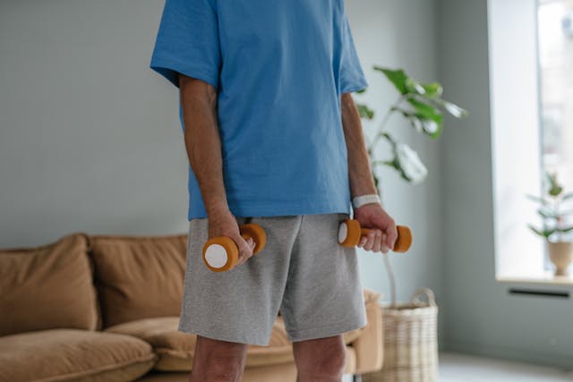
Feeling drained can make even light exercise feel daunting, and this tendency becomes more noticeable as people grow older. The body’s ability to generate energy, recover from exertion, and maintain a steady mood can all shift with age, leaving many individuals feeling that regular activity is too tiring to manage. A recent pilot clinical trial conducted at UT Health San Antonio has identified a potential way to ease some of these barriers. The study explored whether supplementing with branched-chain amino acids (BCAAs) while following a structured exercise routine could help older adults feel less fatigued, improve physical strength, and reduce depressive symptoms. Rather than treating the supplement as a standalone fix, the researchers positioned it as something that might make exercise itself more accessible and rewarding.

The research is grounded in the broader question of how ageing alters the body’s internal systems, particularly those connected to inflammation and metabolism. Jason O’Connor, PhD, a co-author of the study and associate professor in the Department of Pharmacology at The University of Texas at San Antonio, explained that his team has spent years investigating how chronic, low-grade inflammation drives functional decline. Inflammation tends to increase with age and can interfere with both physical and emotional resilience. O’Connor noted that amino acid metabolism has emerged as a key factor in these processes. Amino acids serve as the building blocks of proteins and also help regulate brain chemistry; disruptions in their use and balance may influence energy levels, muscle maintenance, and mood. For older adults already coping with fatigue or reduced mobility, even a slight improvement in metabolic efficiency could make a meaningful difference.

The trial itself was published in the August 2025 issue of Dietetics and ran over eight weeks. Twenty participants, with an average age of about seventy and all living with obesity, took part in a programme combining moderate aerobic exercise and strength training. The study was double-masked and placebo-controlled, meaning that neither participants nor researchers knew who received the BCAA supplement or an inactive placebo. This design helps ensure that neither expectation nor bias affects outcomes. What emerged at the end of the eight weeks was a noticeable contrast between the two groups. While those who exercised without supplementation experienced modest improvements, the group taking BCAAs reported far greater gains. Fatigue decreased by roughly 45%, depressive symptoms declined by about 29%, and there were measurable upticks in strength and endurance.

Understanding why BCAAs might exert these effects requires looking at what they actually are. The three amino acids in this group—leucine, isoleucine, and valine—cannot be produced by the body and must be obtained through food or supplements. They are essential for muscle protein synthesis and recovery, which becomes increasingly relevant as muscle naturally declines with age. Yet the benefits appear to extend beyond muscle tissue. Several lines of research suggest that BCAAs may reduce perceived mental fatigue, perhaps by modulating neurotransmitters involved in alertness and emotional balance. They may also trigger cellular pathways that defend against muscle breakdown. For older adults, these combined effects could translate into feeling more capable during exercise and recovering more comfortably afterwards. In other words, the supplement does not create strength on its own, but it may enable individuals to participate more fully and consistently in the movements that build strength.

Even so, the study’s authors stress the importance of perspective. The improvements seen here, though impressive for a small pilot trial, are not enough on their own to justify universal recommendations. Larger, longer-term studies are needed to confirm the results, determine optimal dosages, and explore how differences in age, health status, or lifestyle may influence outcomes. O’Connor and his research team are already organising further trials, including studies involving ageing veterans. They are also pursuing more detailed inquiries into how amino acid metabolism interacts with inflammation levels, muscle adaptation, and mood regulation. The long-term goal is a clearer, more personalised understanding of how nutrition can support healthy ageing, rather than a one-size-fits-all prescription.

Another crucial factor to consider is that supplements interact differently with different bodies. Although BCAAs are widely available, they can affect medical conditions or medications, especially in older adults managing complex health profiles. This is why the researchers caution that anyone considering adding BCAAs to their routine should first consult a healthcare professional. Supplements can be helpful, but they are most effective when introduced with guidance, context, and awareness of the individual’s overall health. The real value of the study’s findings lies not in encouraging people to rush out and buy supplements, but in showing that there may be supportive strategies that help older adults stay active and feel better while doing so.

Ultimately, this research opens the door to a hopeful message: maintaining activity in later life need not feel like fighting against the body’s limits. With thoughtful support—whether nutritional, behavioural, or therapeutic—it may be possible to feel less exhausted and more empowered. Exercise remains one of the strongest contributors to physical and emotional wellbeing, particularly in ageing. Suppose BCAA supplementation can make the path toward regular activity smoother, more manageable, and more uplifting for individuals who struggle with fatigue or low motivation. In that case, it represents not a shortcut but a gentle nudge toward strength, resilience, and renewed confidence.

More information: Ronna Robbins et al, Branched-Chain Amino Acids Combined with Exercise Improves Physical Function and Quality of Life in Older Adults: Results from a Pilot Randomized Controlled Trial, Dietetics. DOI: 10.3390/dietetics4030032

Journal information: Dietetics Provided by The University of Texas at San Antonio Health Science Center

Why Age Matters in Vaccine Response and What Can Enhance Protection

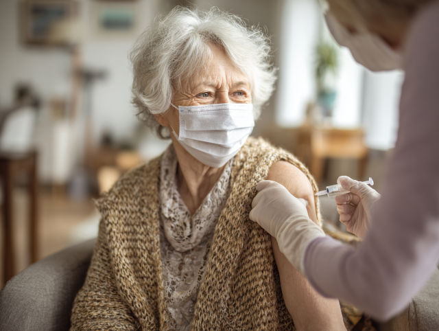
As flu season approaches and vaccination campaigns begin once again, a long-standing challenge comes back into focus: vaccines often do not work as effectively for older adults, particularly those aged 65 and above. Scientists at the Allen Institute have been working to understand why this happens, and their findings are offering new ways to improve vaccine effectiveness later in life. Instead of viewing weaker immune responses as an inevitable decline, the research suggests that the immune system changes in patterned, predictable ways as we age, opening the possibility of targeted solutions that could help vaccines work better for everyone.

The study, published in Nature, is the largest of its kind and focuses on T cells, which play a central role in coordinating immune responses. These cells help B cells produce antibodies that fight viruses and make vaccines more effective. For a long time, researchers believed that chronic inflammation or accumulated health conditions were the leading causes of reduced immune responses in older adults. However, the new findings indicate that, even in healthy ageing, T cells undergo a significant functional shift. This change is not random or simply damage from illness, but rather a fundamental and universal feature of the ageing immune system.

Researchers found that memory T cells in older individuals shift toward a “Th2-like” state, altering the signals they send to B cells. As a result, even when a vaccine delivers the correct viral material, the immune system may struggle to mount a strong antibody response. Lead author Claire Gustafson, PhD, noted that these findings challenge assumptions about the role of inflammation, suggesting that inflammation and immune ageing are not as closely linked as once believed. Instead, immune cells behave differently as we grow older, shaping how effectively the body can respond to vaccines such as the annual flu shot or COVID-19 boosters.

This insight has important implications for future vaccine design. Instead of relying on a single vaccine formulation for people of all ages, researchers may be able to tailor vaccines to account for age-related immune changes. Doctors might one day evaluate a person’s immune profile to predict how well they will respond to a vaccine, allowing for more personalised immunisation strategies. Scientists are also exploring whether immune cells could be adjusted or “retrained” before vaccination. Technologies such as CRISPR, already used in therapies like CAR-T for cancer, could in the future be applied to reprogramme ageing immune cells so they respond more like younger ones.

The conclusions of this study are based on extensive data. Scientists worked with more than 90 healthy adults aged 25 to 65 over 2 years, using advanced techniques such as single-cell RNA sequencing and proteomics to observe how immune cells changed. They then created the Human Immune Health Atlas, an extensive online resource that maps seventy-one types of immune cells across age groups. By analysing more than 16 million immune cells from adults aged 90 and older, the team assembled an unprecedented picture of how immune ageing unfolds. This resource is freely available and is already expected to support future research worldwide.

The significance of this work extends beyond seasonal vaccines. It provides a clearer understanding of how the immune system changes across the lifespan and offers concrete targets for maintaining immune health as we age. As researchers continue exploring this dataset, they hope to uncover further insights that may reshape how medicine supports older adults in staying healthy, resilient, and protected.

More information: Claire Gustafson et al, Multi-omic profiling reveals age-related immune dynamics in healthy adults, Nature. DOI: 10.1038/s41586-025-09686-5

Journal information: Nature Provided by Allen Institute

Cutting Back on Sleep Medication May Enhance Well-Being and Extend Lifespan in Older Adults

Millions of older adults in the United States struggle with chronic insomnia, and many rely on prescription sleep medications to cope. These drugs, including benzodiazepines and Z-drugs such as Ambien, are often taken nightly and sometimes for far longer than medical guidelines recommend. While they can bring short-term relief by helping people fall asleep more easily, they also carry well-documented risks for older adults. These risks include increased likelihood of falls, fractures, memory problems, and physical dependence on the medication. Despite warnings from leading medical societies, the use of these drugs in older age has remained widespread, in part because sleep problems are so persistent and distressing. Older adults may feel they have no other workable option, and doctors sometimes continue prescriptions to avoid causing withdrawal symptoms or renewed insomnia, which can make it seem as though the medication is still necessary.

A recent study conducted by the USC Schaeffer Center for Health Policy & Economics suggests that reducing the use of sleep medications among older adults could lead to significant improvements in both health and life expectancy. The researchers used a detailed simulation model to examine what would happen if people aged fifty and older stopped taking these drugs. Their findings indicated that avoiding these medications could reduce the number of falls in later life by around 8.5 per cent and lower the risk of cognitive impairment by just over 2 per cent. These changes might sound modest on an individual level, but when applied to millions of people across the country, their impact becomes substantial. The researchers also estimated that older adults could gain an average of 1.3 months of additional life, totalling more than a million extra years of life across the population. Most importantly, this added time would be spent in relatively good health rather than simply extending years of frailty.

Sleep medication use is widespread among adults over fifty, with more than fifteen million people in this age group taking prescribed sleep aids. The prevalence tends to increase with age and is exceptionally high among women and white adults. While insomnia itself can be harmful and has been linked to depression, heart disease, and cognitive decline, the long-term use of medication is not proving to be the best answer for many individuals. Part of the difficulty is that withdrawal from these medications can be uncomfortable, causing anxiety and a return of sleeplessness that feels worse than before. This can create confusion for both patients and physicians, who may interpret the withdrawal symptoms as evidence that the medication is still needed. As a result, the prescription continues, even though it may no longer be providing true benefit, and may instead be contributing to further health challenges.

The economic consequences of reducing medication use are also notable. According to the study, avoiding sleep drugs in older adults would lead to an average lifetime savings of around $6,600 per person and approximately $101 billion nationally. These savings stem largely from improvements in health and quality of life, including avoiding falls that lead to hospitalisation and reducing the need for nursing home care. Older adults who fall and break a bone often face long recovery periods, reduced mobility, and, in some cases, a permanent loss of independence. By lowering the odds of these events, reducing the use of sleep medication could help more older adults maintain their daily routines, physical stability, and sense of autonomy for longer. In practical terms, this means fewer medical emergencies, fewer families facing sudden care needs, and a healthcare system less burdened by preventable injuries and complications.

One of the most promising alternatives to prescription sleep medication is cognitive behavioural therapy for insomnia, commonly known as CBT-I. This approach does not involve drugs but instead focuses on teaching individuals how to change habits and thought patterns that interfere with sleep. CBT-I helps people create more consistent sleep routines, address worries that can keep the mind active at night, and build healthier associations with the sleep environment. Research shows that CBT-I is just as effective as medication in the short term and more effective over time, with none of the harmful side effects. It can be delivered in person, through online sessions, or via structured mobile apps, making it accessible to many people. However, awareness and availability can vary, and many patients are not offered CBT-I as a first-line treatment. Encouraging physicians to recommend it more often could be a key step in reducing medication dependence.

The study also highlights several strategies that could help shift prescribing habits. For example, electronic health record systems could be set up to remind doctors of the potential risks when prescribing sleep medication to older adults, or to prompt them to consider alternative treatments. Clinicians might also receive routine feedback comparing their prescribing rates with those of their peers, encouraging reflection and adjustment. These types of interventions have been used successfully in the past to reduce unnecessary prescriptions for antibiotics and opioids. The goal is not to deny medication to those who genuinely need it, but to ensure that prescriptions are thoughtful, limited in duration, and accompanied by support for safer long-term strategies. The researchers emphasise that insomnia is a real and troubling problem for many older adults, and the aim is not to minimise the distress it causes, but instead to encourage solutions that lead to lasting improvements in well-being rather than continued risk.

In essence, reducing the use of prescription sleep medications among older adults represents a shift toward healthier aging. It encourages approaches that prioritise safety, independence, and cognitive clarity. It also acknowledges that sleep is not merely a medical issue but one deeply connected to emotional, social, and lifestyle factors. Helping older adults achieve better sleep without relying on drugs requires patience, guidance, and compassionate support. Yet the potential rewards are considerable: improved daily functioning, reduced risk of injury, enhanced mental sharpness, and a stronger sense of control over one’s health. By encouraging safer sleep practices and broadening access to effective non-drug treatments like CBT-I, both healthcare providers and patients can work together to create more sustainable and empowering approaches to sleep in later life.

More information: Hanke Heun-Johnson et al, Lifetime burden of prescription medication for insomnia in middle-aged and older adults in the US: a microsimulation study, The Lancet Regional Health – Americas. DOI: 10.1016/j.lana.2025.101284

Journal information: The Lancet Regional Health – Americas Provided by University of Southern California

High Intake of Ultra-Processed Foods Associated with Reduced Physical Function in Later Life

A recent study published in Nutrients examined whether eating large amounts of ultra-processed foods influences frailty and physical function in middle-aged and older adults. The researchers found that higher intake of these foods did not directly increase the likelihood of developing frailty, a condition marked by weakness, fatigue and reduced resilience. However, the study showed that consuming more ultra-processed foods was associated with small but noticeable declines in physical performance over time. These changes were subtle, yet meaningful enough to raise concerns about how diet shapes mobility as people age.

The research drew on data from 2,547 adults taking part in the Framingham Offspring Cohort, a long-term study that has followed participants for decades. Over an average of nearly 11 years, around 9 per cent of the participants developed frailty. Despite this, statistical analysis did not show a direct relationship between ultra-processed food intake and the onset of frailty. The results suggest that frailty is influenced by many different factors, including genetics, physical activity levels and overall health history, which may make it challenging to isolate diet as a single cause.

However, when the researchers examined measures of physical function more closely, a more precise pattern emerged. Each additional daily serving of ultra-processed food was associated with a minimal decline in walking speed each year for both men and women. In men, each extra serving was also associated with a slight reduction in grip strength over time. Walking speed and grip strength are both widely used indicators of physical capability in ageing research, as they reflect balance, muscle strength and overall bodily resilience. Even slight declines in these measures can accumulate and eventually affect daily activities.

Shivani Sahni, PhD, a senior author on the study, explained that the findings highlight a more gradual form of impact. Ultra-processed foods may not directly cause frailty, but their nutritional quality may contribute to slower, quieter declines in physical strength and mobility. Such foods often contain high levels of added sugars, unhealthy fats and sodium, while providing fewer nutrients needed to maintain muscle and bone health. Over many years, this imbalance may wear down the body’s ability to move with ease and confidence.

Lead author Elsa M. Konieczynski, MS, emphasised the importance of considering diet as part of ageing well. She noted that choosing whole and minimally processed foods may help preserve physical function and support independence later in life. These foods supply fibre, vitamins, minerals and protein in forms that are more readily used by the body to sustain muscle strength and cellular repair. Shifting everyday eating habits does not require strict avoidance of convenience foods, but rather a mindful approach to balance and nourishment.

The study underscores the idea that healthy ageing is shaped by gradual, long-term patterns rather than sudden changes. While the link between ultra-processed foods and frailty was not direct, the observed declines in gait and strength suggest that diet still plays an important role in how the body ages. These findings add to growing evidence that diets richer in whole foods can support mobility, vitality and independence. For individuals and healthcare providers alike, such research reinforces the value of dietary choices made not just for immediate satisfaction, but for the ability to live actively and confidently in later life.

More information: Elsa M. Konieczynski et al, Ultra-Processed Food and Frailty: Evidence from a Prospective Cohort Study and Implications for Future Research, Nutrients. DOI: 10.3390/nu17162631

Journal information: Nutrients Provided by Hebrew SeniorLife Hinda and Arthur Marcus Institute for Aging Research

Study reveals many Americans misunderstand the link between alcohol consumption and cancer risk

Researchers at The University of Texas MD Anderson Cancer Center have reported that many people in the United States still do not realise that drinking alcohol increases the risk of developing cancer. This is notable because the scientific evidence linking alcohol to several types of cancer is well-established and has been acknowledged by major health organisations for years. Yet the study found that more than half of American adults surveyed did not recognise this connection. The research raises concerns about how health information is shared and understood, and why certain risks remain poorly acknowledged in everyday life. Suppose people are unaware that a behaviour poses a potential danger. In that case, they are unlikely to adjust it, which means the effects of misinformation or simple lack of awareness can have real consequences for public health.

The study, published in JAMA Oncology, noted that around 52.9 per cent of adults did not know that alcohol affects cancer risk. In comparison, only 37.1 per cent correctly recognised that drinking increases the likelihood of developing cancer. A tiny minority, roughly one per cent, believed that alcohol might actually decrease cancer risk. Another striking detail from the findings was that people who had recently consumed alcohol were more likely to think that it did not affect cancer risk. This suggests that personal habits may influence how individuals interpret scientific information, sometimes in ways that protect them from discomfort or guilt. When a behaviour is socially common, culturally normalised, and frequently associated with pleasure or relaxation, it can be easier for individuals to discount warnings, even when the evidence is clear.

Lead author Sanjay Shete, PhD, pointed out that beliefs are central to health decision-making. If someone does not believe that alcohol contributes to cancer, they are unlikely to feel motivated to reduce how much they drink. Shete emphasised that addressing such misunderstandings is essential to encouraging healthier choices at the population level. The argument is not merely about persuading people to stop drinking entirely, but rather ensuring they know the risks clearly enough to make informed decisions. Alcohol marketing and social culture tend to focus on enjoyment, sociability, and lifestyle, with very little attention given to long-term health consequences. This cultural framing can complicate efforts to communicate medical risks effectively, even when the science is clear and consistent.

The study also highlighted differences in awareness among demographic groups. Individuals with lower levels of educational attainment, current cigarette smokers, and those who identified as Black were less likely to know about the cancer risks associated with alcohol. Additionally, people who did not believe that cancer can be prevented were more inclined to think alcohol did not influence cancer risk. These findings underline that public health messaging must consider social context, access to information, and structural inequalities. Presenting data alone is not always sufficient to change beliefs. Messages may need to be communicated in ways that are culturally relevant, accessible, and sensitive to how different communities receive and interpret health guidance.

Alcohol is classified by the World Health Organization as a Group 1 carcinogen, placing it in the same category as substances such as tobacco, asbestos, and certain forms of radiation. This classification indicates that there is substantial and conclusive evidence that alcohol can cause cancer. Drinking has been linked to cancers of the breast, liver, colon, throat, mouth, and oesophagus, among others. According to the National Institutes of Health, about 5.5 per cent of all new cancer cases and 5.8 per cent of cancer-related deaths can be attributed to alcohol consumption. These are not small numbers, and they illustrate why awareness matters. If more people understood that routine drinking has a measurable impact on cancer risk, it could influence how often and how much they choose to drink. This is especially relevant given recent shifts in guidelines that now encourage lower levels of alcohol consumption for health protection.

The researchers based their conclusions on responses from nearly 7,000 adults who took part in the 2024 Health Information National Trends Survey. Participants were asked a direct question: how they believed drinking alcohol affects cancer risk. They could answer that it increases risk, decreases risk, has no effect, or that they did not know. The varied responses reflected not only a lack of information but also the influence of personal experience, social environment, and cultural messaging. In short, the study reveals a significant divide between scientific knowledge and public understanding. Bridging this divide will require clear communication, better education, and more consistent public health messaging. By increasing awareness and correcting misunderstandings, it may be possible to reduce preventable cancer cases and encourage healthier approaches to alcohol consumption across society.

More information: Joël Fokom Domgue et al, Beliefs About the Effect of Alcohol Use on Cancer Risk in the US Adult Population, JAMA Oncology. DOI: 10.1001/jamaoncol.2025.4472

Journal information: JAMA Oncology Provided by University of Texas M. D. Anderson Cancer Center

Extent of Plant-Food Processing as a Determinant of Heart Health

Many earlier studies have suggested that eating a large amount of ultra-processed foods is linked to an increased likelihood of developing cardiovascular disease. At the same time, other research has shown that diets rich in plant-based foods, when of good nutritional quality, may help lower this risk. However, simply labelling a diet as “plant-based” does not always mean it is healthy, because plant-derived foods can vary widely in how they are prepared and processed. A diet full of vegetables, whole grains, legumes, fruits, nuts, and seeds in their natural or lightly processed forms is not the same as a diet high in packaged snacks, sweetened drinks, and ready-made meals, even if all of these are technically made from plants. As plant-based eating becomes more popular, it has become increasingly important to understand what “plant-based” really means in practice, and how different types of plant-derived foods affect the body.

A team of researchers from INRAE, Inserm, Université Sorbonne Paris Nord, and Cnam examined this question in depth by analysing not only whether people ate more plant-based or animal-based foods, but also the nutritional value of the foods and the level of industrial processing involved. Nutritional quality refers to how foods are composed in terms of fat, sugar, salt, fibre, and key vitamins and minerals. The level of processing, on the other hand, refers to how much the food has been altered from its original form through factory preparation, use of additives, or refinement. By combining these two considerations—quality and processing—the researchers were able to gain a clearer picture of how modern dietary patterns relate to heart health, beyond simple categories such as vegetarian, vegan, or omnivorous.

To conduct this research, the scientists used data from 63,835 adults participating in the long-running NutriNet-Santé study in France. The participants’ diets were recorded through detailed online questionnaires that documented their food and drink consumption over several days. The average follow-up period for monitoring their health outcomes was just over 9 years. For some individuals, the study followed their dietary patterns and cardiovascular health for up to 15 years. With this information, the researchers identified different types of diets not only by the proportion of plant-based versus animal-based foods, but also by the nutritional value of those foods and the degree of processing before consumption.

The results showed that individuals who consumed a high proportion of plant-based foods that were both nutritionally balanced and minimally processed had a significantly lower risk of developing cardiovascular diseases. Specifically, those whose diets emphasised fresh or lightly processed plant foods—such as vegetables, whole fruits, whole grains, legumes, and nuts—had roughly a 40 per cent lower risk of heart disease compared with people whose diets contained fewer such foods and more animal-based products. These findings support existing public health guidance encouraging diets built around whole plant foods that are rich in fibre, antioxidants, and healthy fats, while being relatively low in added sugars, salt, and saturated fats.

However, the study also revealed that eating a diet high in plant-based foods does not automatically offer protection if those foods are ultra-processed. Individuals who ate substantial amounts of plant-based foods that underwent extensive industrial processing did not experience a reduced risk of cardiovascular disease, even when these products appeared nutritionally acceptable on labels. Examples of such foods include pre-packaged soups, industrial breads, ready-made meals, and salads prepared with processed dressings. Ultra-processed foods often contain additives, refined ingredients, and altered food structures that may affect metabolism, digestion, and appetite regulation. This means that, in theory, a plant-based diet may still act in the body in ways that do not support heart health if the foods have been heavily processed during manufacturing.

The study further found that diets high in plant-based foods, both low in nutritional quality and ultra-processed, were associated with an increased risk of cardiovascular disease. This category included sugary cereals, sweetened drinks made from plant extracts, confectionery, flavoured snack foods, and similar items. In these cases, the risk of heart disease increased by about 40 per cent when compared with people consuming minimally processed, nutrient-rich plant foods. These findings highlight an important point for public health messaging: encouraging plant-based eating is beneficial only when the foods promoted are genuinely nutritious and as close as possible to their natural forms. For dietary guidance to be practical, it must consider not only the plant-to-animal ratio in the diet but also the nutritional composition and degree of processing of the foods consumed.

More information: Clémentine Prioux et al, Cardiovascular disease risk and the balance between animal-based and plant-based foods, nutritional quality, and food processing level in the French NutriNet-Santé cohort: a longitudinal observational study, The Lancet. DOI: 10.1016/j.lanepe.2025.101470

Journal information: The Lancet Provided by INRAE – National Research Institute for Agriculture, Food and Environment

Research Reveals How Social and Economic Hardship in Local Communities May Contribute to Higher Dementia Rates

Cambridge researchers have revealed new evidence explaining why living in a socioeconomically disadvantaged neighbourhood may raise a person’s likelihood of developing dementia. Their study suggests that the environment in which someone lives can influence both the condition of the brain’s small blood vessels and the ability to maintain lifestyle habits that protect cognitive health. Rather than dementia risk being shaped only by personal choices or genetics, this research strengthens the argument that broader social conditions play a major role in long-term brain health. The findings, published in Alzheimer’s & Dementia: The Journal of the Alzheimer’s Association, provide further clarity on how midlife deprivation can set the stage for cognitive decline decades later.

Dementia has long been observed to affect people in disadvantaged communities more frequently and at earlier stages. Importantly, this increased risk appears even among individuals who are financially stable or well educated themselves, suggesting that the characteristics of the neighbourhood environment shape health outcomes beyond individual circumstances. Earlier research has also found that people in deprived areas tend to show more signs of structural brain damage, including stress to brain tissue, though the underlying reasons have not been well understood. This new study offers evidence that these effects may be linked to the health of small blood vessels in the brain, which are essential for carrying oxygen and nutrients to the neural systems involved in memory, attention, and decision-making.

The Cambridge team examined data from 585 adults aged 40 to 59 living across the UK and Ireland, who were participating in the PREVENT-Dementia programme, which follows people in midlife to identify early risk factors. Participants were assessed on several measures: the level of neighbourhood deprivation based on their postcode; their performance on cognitive tests; lifestyle risk factors such as sleep quality, physical activity, weight, and blood pressure; and MRI scans to detect any early signs of damage in the brain’s small vessels. By comparing these factors, the researchers were able to explore how environmental disadvantage may influence brain health both directly and indirectly.

The study showed that individuals living in areas with higher unemployment rates, lower incomes, or fewer educational opportunities were more likely to experience difficulties maintaining healthy lifestyle habits. They were more likely to have poor sleep, higher blood pressure, obesity, and lower physical activity levels. All of these are known contributors to dementia risk because they can damage the brain’s vascular system over time. Interestingly, people in more deprived communities tended to drink less alcohol than those in more affluent neighbourhoods, reminding us that health patterns vary across contexts and that public health strategies must be tailored accordingly.

Neighbourhood deprivation was also associated with reduced cognitive performance, particularly in processing speed, spatial awareness, and concentration. These skills are vital for everyday functioning and often decline early in the path toward dementia. The researchers believe this reduced cognitive ability may be tied to the vascular damage seen on brain scans. When small vessels are weakened or obstructed, the brain receives less oxygen and fewer nutrients, which can gradually impair neural function. Crucially, the findings showed that these effects were not simply a matter of individual education levels or personal background. Even people with university degrees and stable employment were affected when they lived in deprived areas, highlighting how the physical and social environment can shape behaviour and health in powerful ways.

The results of this study emphasise that dementia prevention must go beyond encouraging individuals to make healthier lifestyle choices. While personal habits are essential, people can only act within the possibilities their environment allows. If someone lives in a neighbourhood without safe public spaces, access to affordable, healthy food, reliable healthcare, or a sense of security, maintaining good health becomes far more difficult. The researchers argue that meaningful dementia prevention will require policymakers to address the social conditions that limit healthy living. Improving neighbourhood safety, creating accessible exercise spaces, reducing economic inequality, and strengthening community support systems could all help lower dementia risk at a population level. In short, the study reinforces the idea that where we live deeply matters for how our brains age.

More information: Audrey Low et al, Neighborhood deprivation and midlife cognition: Evidence of a modifiable vascular pathway involving health behaviors and cerebral small vessel disease, Alzheimer’s & Dementia. DOI: 10.1002/alz.70756

Journal information: Alzheimer’s & Dementia Provided by University of Cambridge

Fresh study reveals: neurons build ‘air-tunnel’ networks that circulate harmful proteins linked to Alzheimer’s disease

Scientists at Johns Hopkins Medicine have discovered that mammalian brain cells can form microscopic tubes that transport toxic molecules between neurons, functioning like tiny “pneumatic tubes” within the brain. This process, observed in experiments using genetically engineered mice, helps explain how harmful proteins linked to Alzheimer’s disease might spread from one cell to another. The study, funded by the National Institutes of Health and published on 2 October in Science, could change how researchers think about the brain’s internal communication systems and open new directions for treating neurodegenerative disorders.

In their experiments, the researchers found that neurons form slender channels called nanotubes to remove small toxic molecules. One of them is amyloid-beta, a sticky protein that forms plaques that mark Alzheimer’s disease. Hyungbae Kwon, an associate professor of neuroscience at Johns Hopkins, explained that while these tubes help cells expel toxins, they also facilitate their spread. “Cells have to get rid of toxic molecules,” Kwon said, “but by producing a nanotube, they can transmit this toxic molecule to a neighbouring cell. Unfortunately, this also spreads harmful proteins through the brain.”

Using high-powered microscopes and live-cell imaging, the team watched neurons extend long, finger-like projections that connected one cell’s dendrites — the branch-like arms — to another’s. These structures, named dendritic nanotubes, acted as highways for transporting small molecules, calcium, and even toxic proteins. The researchers noted that the shape and flexibility of the tubes enabled them to rapidly transfer information and materials between cells that were not directly touching, revealing a form of communication previously hidden from scientists.

To explore this process further, the team built computer models to simulate how nanotubes contribute to the buildup of amyloid-beta early in Alzheimer’s disease. Their findings showed that the brain may have an extra “connectivity layer” of nanotubes that works alongside synapses — the traditional communication points between neurons. This suggests that the brain’s internal network is more physically linked than previously thought, and that these nanotubes might play a role in both maintaining and disrupting brain health.

The researchers examined brain tissue from healthy mice and from mice genetically modified to develop Alzheimer ‘s-like plaques. They discovered that diseased mice had more nanotubes at 3 months of age, before symptoms appeared, than healthy mice. By six months, the difference between the two groups had lessened. This pattern suggests that nanotube formation increases during the early stages of disease, possibly facilitating the rapid transport of toxic proteins through brain tissue. When the scientists studied human brain samples using publicly available electron microscopy data, they found nanotubes forming between neurons, a pattern similar to that observed in mice, suggesting that this mechanism exists in both humans and mice.

Kwon and his team now plan to test whether other brain cells, such as glial cells, also form nanotubes. They hope to learn whether controlling the creation of these tubes could help prevent or slow diseases like Alzheimer’s. If researchers can find a way to adjust nanotube formation — increasing it to remove waste or reducing it to stop the spread of toxins — they might one day use this discovery to protect brain cells from degeneration. This new understanding of how the brain moves molecules around adds an intriguing piece to the puzzle of how neurodegenerative diseases begin and progress. It offers hope for more targeted, effective treatments in the future.

More information: Hyungbae Kwonet al, Intercellular communication in the brain through a dendritic nanotubular network, Science. DOI: 10.1126/science.adr7403

Journal information: Science Provided by Johns Hopkins Medicine

Guiding Screens and Streams: Grandparents’ Role in Children’s Media Engagement

A new study from Rutgers University has found that grandparents play a far more important role in managing children’s media use than previously recognised. Conducted by Dafna Lemish, a Distinguished Professor of Journalism and Media Studies at Rutgers, along with Galit Nimrod and Nelly Elias from Ben-Gurion University of the Negev, the research explores how grandparents act as mediators in their grandchildren’s digital lives. Published in the Journal of Aging Studies, the study introduces the idea of “secondary mediation,” a process where grandparents use strategies and rules created by parents to guide children’s media habits. The researchers argue that traditional mediation theories should be expanded to include this shared, intergenerational approach to caregiving.

The study highlights that grandparents often follow parental guidelines when supervising television, online content, and games, effectively reinforcing family values around media use. However, this cooperation depends significantly on the relationship between the parents and grandparents, as well as the grandparents’ own familiarity with technology. The authors found that this collaboration strengthens intergenerational bonds, turning media supervision into a shared effort rather than a source of conflict. It also demonstrates that grandparents are not just passive observers in childcare but active partners who help maintain consistency in children’s digital routines.

Professor Lemish explained that much of the research on children’s media consumption has focused mainly on parents, overlooking grandparents who are frequently involved in caregiving. She wanted to know whether grandparents used the same strategies as parents—restricting screen time, discussing content, and co-viewing—or were more relaxed, perhaps “spoiling” their grandchildren. As both a scholar and a grandmother, Lemish was personally drawn to understanding these dynamics. Her findings reveal that grandparents do engage in media mediation, though often in more informal and emotionally supportive ways, reflecting their unique role in family life.

The research found that grandparents can have a positive influence by reducing children’s exposure to harmful content and encouraging media that supports learning and creativity. Lemish noted that grandparents can help counteract adverse effects such as exposure to violence, bullying, or stereotypes, while promoting enjoyment and intellectual growth. This balance of caution and encouragement allows children to benefit from the educational and social advantages of media without overindulgence. Grandparents’ wisdom and experience, the study suggests, can help young children navigate digital spaces more safely and thoughtfully.

A key recommendation from the researchers is that parents and grandparents should communicate openly about media rules and expectations. When caregivers share similar values and guidelines, children are less likely to experience confusion or tension. Lemish advised families to discuss their perspectives on media, agree on limits for screen time, and decide together when exceptions might be acceptable. This cooperative approach ensures that children receive consistent messages about using media responsibly while still allowing for flexibility and enjoyment.

To reach their conclusions, the researchers surveyed 267 pairs of grandmothers and mothers of children aged four to eight. They discovered that grandmothers were more confident in mediating noninteractive media, such as films or television, than interactive media, such as games. Factors such as education, time spent caregiving, and comfort with technology influenced how they guided their grandchildren. Overall, the study provides some of the first detailed insights into how grandparents shape children’s media habits. It portrays them as vital partners in helping families find balance in an age where screens are a constant presence in everyday life.

More information: Galit Nimrod et al, Secondary mediation of children’s media use: An exploration of mothers-grandmothers’ mediation dynamic, Journal of Aging Studies. DOI: 10.1016/j.jaging.2025.101357

Journal information: Journal of Aging Studies Provided by Rutgers University

Alterations in brain structure may reveal early indicators of dementia

A new study from the University of California, Irvine’s Center for the Neurobiology of Learning and Memory has found that ageing changes the brain’s shape in ways that can be measured and linked to mental decline. Rather than focusing only on the size of individual regions, researchers used a new analytic method to track how the brain’s overall form shifts and distorts over time. Their findings reveal that these geometric changes could provide early warning signs of cognitive impairment.

The research, published in Nature Communications and supported by the National Institute on Aging, showed that the brain’s shape changes systematically with age. The study’s senior author, Dr Niels Janssen of Universidad de La Laguna and UC Irvine, explained that while most studies focus on tissue loss, this one uncovered how the brain’s entire structure moves and warps in patterns tied to memory and reasoning decline. These shifts, he noted, could be a powerful new way to gauge brain health.

Using more than 2,600 brain scans from adults aged 30 to 97, the researchers found that some areas of the brain expand outward while others shrink inward with age. Specifically, the inferior and anterior areas of the brain tended to bulge, whereas the superior and posterior regions compressed. This uneven reshaping was most pronounced in people showing signs of cognitive decline, and those with greater posterior compression often performed worse on reasoning tasks. The same pattern appeared across multiple datasets, confirming the reliability of these changes as a hallmark of ageing.

One particularly intriguing finding involves the entorhinal cortex, a small but vital memory centre in the brain. The study suggests that as the brain reshapes over time, this region may be physically pushed closer to the hard base of the skull. This compression could make it more vulnerable to damage, particularly from tau, the toxic protein associated with Alzheimer’s disease. The research team proposes that these mechanical stresses may partly explain why this region is often one of the first affected in Alzheimer’s – a new way of thinking about how the disease begins.

Dr Michael Yassa, co-author and director of UC Irvine’s centre, said this discovery could change how scientists view early Alzheimer’s detection. He explained that if brain shape changes gradually increase pressure on fragile regions, it could trigger the cascade of damage that leads to dementia. Identifying these geometric markers early could one day help detect risk years before symptoms appear, providing a valuable window for intervention.

The study, a collaboration between UC Irvine and Universidad de La Laguna, highlights the promise of using brain geometry as a new diagnostic tool. As Dr Janssen noted, this research goes beyond simply measuring brain shrinkage—it shows how the brain’s architecture evolves with age and predicts who may be at higher risk for cognitive decline. By uncovering these patterns, scientists may be closer to understanding the physical forces that shape the ageing brain and its connection to diseases like Alzheimer’s.

More information: Yuritza Y. Escalante et al, Age-related constraints on the spatialgeometry of the brain, Nature Communications. DOI: 0.1038/s41467-025-63628-3

Journal information: Nature Communications Provided by University of California – Irvine

Gene Responsible for Familial Vision Loss Found

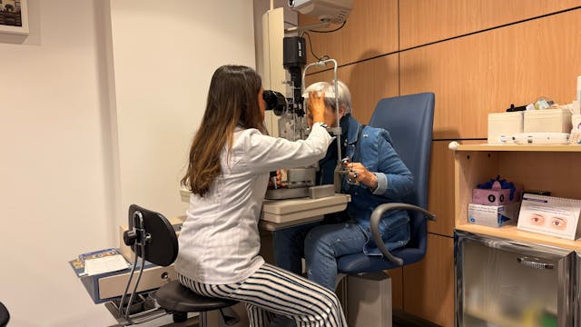
A team of scientists from the Medical University of Vienna and the Medical University of Graz has uncovered a new genetic cause of hereditary optic atrophy. This degenerative disease leads to gradual vision loss. Their findings, recently published in Genetics in Medicine, offer new opportunities for diagnosis and research into the underlying biological processes responsible for the condition. This discovery represents a significant step forward in understanding why some families are affected by inherited vision loss.

The research began with the study of a large Austrian family in which seven members across three generations suffered from optic atrophy. Genome-wide sequencing revealed a previously unknown mutation in the PPIB gene, which encodes an enzyme that helps proteins fold correctly and removes defective ones. In cells from affected individuals, this mutation was found to disrupt mitochondrial function—the “power plants” of cells that provide the energy needed for healthy vision.

Further analysis of existing genetic databases led the team to identify 12 additional individuals from 8 unrelated families who shared the same PPIB mutation, confirming its link to the disease. According to study leader Professor Wolfgang M. Schmidt of MedUni Vienna, the discovery establishes PPIB as a new optic atrophy gene. His colleague, Dr Thomas P. Georgi of MedUni Graz, highlighted that this finding allows for more precise genetic diagnoses and better support for affected families, helping clinicians offer more accurate advice and care.

Optic atrophy damages the optic nerve, which transmits signals from the retina to the brain. It causes a gradual loss of visual clarity, reduced colour perception, and central blind spots. Although around twenty genetic forms of optic atrophy are currently known—most related to mitochondrial dysfunction—about 60 per cent of patients still lack a confirmed genetic explanation. Identifying PPIB helps fill this gap, giving researchers a new clue to explore and patients a more accurate understanding of their condition.

Beyond its relevance to optic atrophy, the discovery of the PPIB mutation could shed light on other diseases linked to mitochondrial damage and protein misfolding. As genome sequencing becomes increasingly available, testing for PPIB variants may soon become part of standard genetic screenings for unexplained vision loss. This breakthrough, therefore, not only advances scientific knowledge but also holds real promise for improving diagnosis, care, and future treatment of inherited eye diseases.

More information: Thomas P. Georgi et al, A recurrent missense variant in the PPIB gene encoding peptidylprolyl isomerase B underlies adult-onset autosomal
dominant optic atrophy, Genetics in Medicine. DOI: 10.1016/j.gim.2025.101595

Journal information: Genetics in Medicine Provided by Medical University of Vienna