Monthly Archives: August 2026

Different Organs, Different Ageing Clocks: A Blood Test Could Reveal Yours

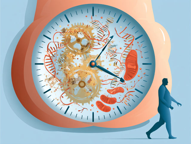
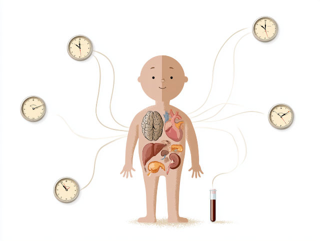
Researchers have developed artificial intelligence (AI)-based “tissue clocks” that can estimate the biological age of human organs from microscopic tissue images. Scientists at the CeMM Research Center for Molecular Medicine of the Austrian Academy of Sciences and the Ludwig Boltzmann Institute for Network Medicine (LBI-NetMed) at the University of Vienna analysed more than 25,000 tissue samples across 40 tissue types. Their findings, published in Nature Medicine, suggest that organs age at different rates and that some of these ageing patterns may even be detectable through blood samples.

People of the same chronological age can differ greatly in how old they appear or how well their bodies function. This raises an important question: do individual organs also age at different speeds? Led by CeMM and LBI-NetMed Principal Investigator André Rendeiro, the research team used AI to investigate how the microscopic architecture of human tissues changes with age, rather than focusing only on molecular markers such as DNA methylation or gene expression.

The researchers used data from the Genotype-Tissue Expression Project (GTEx), which collected samples from 983 individuals across 40 tissue types, including the brain, heart, lungs, pancreas, skin, and intestine. In total, they analysed 25,712 high-resolution tissue images, representing approximately 480 million individual image tiles, using advanced computer vision models.

The analysis showed that age was the strongest factor associated with changes in tissue appearance across all 40 tissue types. Building on this finding, the researchers developed “tissue clocks” capable of estimating biological age independently for different organs. These models predicted age with an average error of just 4.9 years. The estimated biological ages were also associated with established indicators of ageing, including shorter telomeres, tissue abnormalities, and the number of chronic diseases an individual had.

Importantly, the study found that organs do not follow the same ageing schedule. The lungs, kidneys, pancreas, and adrenal glands showed signs of accelerated ageing as early as ages 20 to 40, while other tissues followed more complex patterns later in life. The uterus showed a particularly notable change around menopause. Health conditions were also associated with organ-specific ageing: kidney failure was linked with accelerated ageing signals across several tissues, while diabetes had especially pronounced effects in the pancreas.

The tissue clocks also identified individuals whose organs appeared biologically older than their chronological age. According to the researchers, AI can detect subtle structural changes that may be difficult for the human eye to recognise, allowing ageing to be viewed as a process of architectural remodelling within tissues rather than simply the accumulation of molecular changes.

Because obtaining tissue samples is often invasive or impractical, the researchers investigated whether the same organ-ageing signals could be detected in blood. By linking blood-based gene expression with tissue age estimates from the same individuals, they developed predictors capable of estimating tissue-specific biological ageing using blood samples alone. This could potentially provide a much less invasive way to assess how individual organs are ageing.

The blood-based models detected ageing patterns associated with conditions including Alzheimer’s disease, Crohn’s disease, cystic fibrosis, vasculitis, diabetes, and stroke. Alzheimer’s disease produced its strongest ageing signal in the brain, while Crohn’s disease showed accelerated ageing across the gastrointestinal tract. Although further research is needed before these methods could become routine clinical tests, the findings suggest that combining AI, tissue imaging, and blood analysis could eventually help monitor organ health, detect disease-related changes earlier, and provide a more personalised picture of biological ageing.

More information: Ernesto Abila et al, Histological aging signatures for monitoring tissue-specific aging and disease, Nature Medicine. DOI: 10.1038/s41591-026-04566-5

Journal information: Nature Medicine Provided by CeMM Research Center for Molecular Medicine of the Austrian Academy of Sciences

Research Shows Waist Size Can Help Predict Health Risks

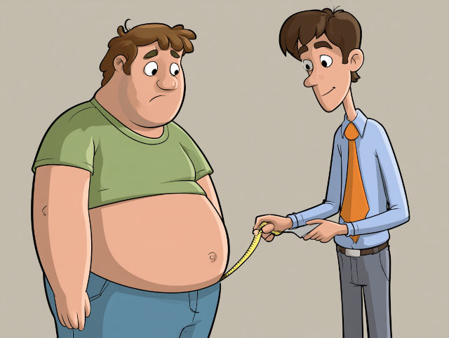
Measuring waist size may be one of the simplest and most effective ways to identify obesity-related health risks, according to a study by Rutgers Health researchers. The findings suggest that waist circumference can provide useful information about unhealthy levels of body fat and may help identify people at increased risk of chronic diseases.

The study, published in JAMA Network Open, found that combining body mass index (BMI) with waist circumference—or using waist circumference alone—performed about as well as a newly proposed definition of obesity that relies on several body measurements. This suggests that a relatively simple measurement may offer much of the information needed for obesity screening.

Researchers analysed data from 1,900 U.S. adults aged 20 to 59 who participated in the National Health and Nutrition Examination Survey. They compared several methods of diagnosing obesity against a highly accurate measure of body fat to determine how well each approach identified people with excess body fat.

The researchers also examined a recently proposed obesity framework developed by a multidisciplinary group of independent experts. The framework incorporates BMI, waist circumference, waist-to-height ratio and waist-to-hip ratio. Although this approach identified more people with excess body fat, the researchers found that simpler methods achieved similar accuracy.

In particular, using either BMI or waist circumference provided a good balance between correctly identifying people with obesity and avoiding false-positive diagnoses. Waist circumference alone performed nearly as well. This is important because waist measurement is relatively easy to perform and can be incorporated into routine health assessments without requiring specialised equipment.

“Pay attention to the need to loosen your belt or get bigger pants,” said lead author Aayush Visaria, a Rutgers Health faculty member. Although clothing size is not a substitute for properly measuring waist circumference, Visaria said changes in clothing fit may provide an early indication of unhealthy amounts of body fat. Monitoring both waist size and weight could therefore help identify health risks earlier.

The study also highlighted some limitations of relying on BMI alone. Researchers found that BMI missed a substantial number of people who met body-fat criteria for obesity. BMI is based on weight relative to height and does not fully account for differences in body composition or where fat is distributed. Waist circumference can provide additional information about abdominal fat, which is associated with risks such as diabetes, heart disease and stroke.

The findings could help shape future recommendations for obesity screening and encourage wider use of waist measurements in routine health care. The researchers noted that some measurements included in newer obesity frameworks, such as hip circumference, are rarely collected in everyday clinical practice. Waist circumference, by comparison, is increasingly recognised in clinical guidelines and is practical for both clinicians and patients. As Visaria explained, multiple complex measurements may not always be necessary: a simple waist measurement can provide valuable information for identifying obesity-related health risks.

More information: Aayush Visaria et al, Lancet Definition vs Other Criteria for Obesity Diagnosis in Adults, JAMA Network Open. DOI: 10.1001/jamanetworkopen.2026.27738

Journal information: JAMA Network Open Provided by Rutgers University

PCB Contamination Detected in Chinese Tea, but Consumer Health Risks Remain Low

Tea is enjoyed by millions of people every day, but tea leaves can also accumulate traces of persistent environmental pollutants during cultivation and production. A new nationwide study provides one of the most comprehensive assessments to date of polychlorinated biphenyls (PCBs) in commercial tea produced across China.

Researchers analysed 192 tea samples covering all six major tea categories and 16 Chinese provinces, testing for 18 PCB compounds, including six indicator PCBs and 12 dioxin-like PCBs. Although PCBs were detected in all samples, the health risk assessment found that exposure from drinking Chinese tea is unlikely to pose an appreciable health risk to consumers.

“Tea leaves have a long journey from the field to the cup, and contaminants can potentially enter at several stages,” said corresponding author Wenfeng Zhou. “Our study provides a national baseline that helps us understand where PCBs occur in tea, how processing may influence their levels, and what those concentrations mean for consumers.”

Total concentrations of the 18 PCBs ranged from 9.36 to 4,490 picograms per gram (pg/g) of dry tea, with substantial differences among tea types. Dark tea had the highest average concentration at 549 pg/g, followed by oolong tea at 241 pg/g, while yellow tea recorded the lowest average concentration at 93.3 pg/g.

Geographic variations were also observed. Tea produced in Guangdong had the highest average PCB concentration at 505 pg/g, followed by Yunnan, Chongqing, Guangxi and Shaanxi. Researchers suggest these differences could reflect local environmental contamination, historical industrial activities and variations in tea production practices. Analysis of the chemical profiles showed that lower-chlorinated PCBs were dominant, with CB-28 and CB-52 contributing substantially to the overall PCB profile.

The researchers also examined whether tea processing itself could affect PCB concentrations by comparing fresh green tea leaves with tea made from the same batch. Total PCB levels increased from 51.2 to 63.7 pg/g after processing, largely because of increases in indicator PCBs. However, the difference was not statistically significant, suggesting that processing was not a major source of PCB contamination in this experiment.

For consumers, the health risk assessment offered reassuring results. Using World Health Organization toxic equivalency factors and typical tea consumption rates, researchers estimated exposure to dioxin-like PCBs. Green tea produced the highest estimated weekly intake because it is consumed in larger quantities, but estimated exposure remained well below established tolerable intake thresholds.

The authors caution that their assessment considers PCB exposure from tea alone rather than a person’s total dietary exposure. Because PCBs persist in the environment and can accumulate through food chains, continued monitoring is important. The study establishes a national baseline for PCBs in Chinese commercial tea, providing evidence that could strengthen food safety surveillance and help identify opportunities to reduce contamination throughout the tea production chain.

More information: Xingyi Wu et al, Profiling of polychlorinated biphenyls in Chinese tea: national distribution, variety-specific variations, and the impact of processing, New Contaminants. DOI: 10.48130/newcontam-0026-0019

Journal information: New Contaminants Provided by Shenyang Agricultural University Collaborative Journals

Socioeconomic Disadvantage Is Associated with Greater Physical Decline with Age

In England and Canada, socioeconomic disadvantage is associated with poorer physical function and greater declines as people age, according to a study published August 13 in the open-access journal PLOS Medicine. The study was led by Stephanie Schrempft of Geneva University Hospitals, Switzerland, and colleagues.

Physical function naturally declines with age, but the rate of decline varies considerably between individuals and populations. Wealth is known to influence health and disability, yet few studies have directly compared patterns of functional ageing across countries. Such comparisons can help researchers understand how social policies and broader structural factors may contribute to inequalities in healthy ageing.

To investigate these differences, researchers analysed data from two major longitudinal studies: 8,511 participants in the English Longitudinal Study of Ageing and 22,605 participants in the Canadian Longitudinal Study on Aging. Participants were between 50 and 85 years old. The researchers examined wealth in relation to walking speed, grip strength, lung function, hearing, mobility, and difficulties performing daily activities such as dressing, bathing and eating.

People with less wealth generally had poorer physical function and experienced greater declines in mobility and their ability to perform daily activities as they aged. Importantly, socioeconomic inequalities in functional ageing were substantially greater in England than in Canada. These differences remained even after researchers accounted for chronic health conditions, weight status and behaviours such as smoking.

The size of the wealth gap was particularly striking for walking speed. In England, the least wealthy 60-year-olds had walking speeds comparable to those of the wealthiest 75-year-olds—an apparent functional ageing difference of approximately 15 years. In Canada, the corresponding difference was about nine years, suggesting meaningful differences in socioeconomic inequalities between the two countries.

The study also identified important differences between women and men. Women with the least wealth experienced greater difficulties with mobility and daily activities than men in the same socioeconomic group, although they had better lung function. These findings demonstrate that socioeconomic disadvantage does not affect everyone in the same way and highlight the value of considering both wealth and sex when studying healthy ageing.

Senior author Silvia Stringhini said the researchers were particularly struck by the substantially larger wealth gaps in England than in Canada. Despite both countries having universal healthcare and similar levels of income inequality, the differences in functional ageing were considerable. She noted that ongoing cross-country research will investigate the structural factors that may explain why these inequalities differ between countries.

First author Stephanie Schrempft emphasised that the findings go beyond showing that people with less wealth have poorer health on average. By following adults over time in England and Canada, the researchers found that socioeconomic disadvantage was also associated with a faster loss of mobility and physical independence with age. Although the study was observational and could not establish causation or fully account for factors such as childhood circumstances and access to private healthcare, the authors suggest that healthy-ageing policies should prioritise socioeconomically disadvantaged populations and address the broader structural conditions underlying these inequalities.

More information: Stephanie Schrempft et al, Socioeconomic inequalities in functional ageing trajectories in England and Canada: A comparative longitudinal cohort study, PLOS Medicine. DOI: 10.1371/journal.pmed.1004833

Journal information: PLOS Medicine Provided by PLOS

Early Life Adversity Leaves a Lasting Molecular Mark Across the Body

The experiences we face early in life may leave lasting marks on our health, with effects that can echo across decades and throughout the body. A new study published in Science provides compelling molecular evidence that early life adversity can leave a lasting, system-wide signature on the epigenome. This biological layer helps regulate gene activity.

Led by researchers at Arizona State University and Vanderbilt University, along with collaborating institutions, the study examined 237 free-ranging rhesus macaques living on Cayo Santiago, a 38-acre island off Puerto Rico. The macaques have been followed throughout their lives, providing researchers with unusually detailed records of their experiences. The team paired these life histories with genomic data from 12 tissues collected in adulthood.

Researchers focused on DNA methylation, one of the most extensively studied epigenetic markers of ageing. Patterns of DNA methylation can be used to create “epigenetic clocks” that estimate both chronological age and biological age. The researchers developed highly precise tissue-specific clocks capable of predicting the macaques’ ages to within approximately one year.

The results revealed that ageing does not occur uniformly throughout the body. Age-related DNA methylation patterns differed substantially among tissues, with some, including the thymus and pituitary gland, showing particularly great and distinctive changes. Blood—the tissue most commonly examined in human studies—captured only part of this complex picture. Despite these differences, individuals who appeared biologically older in one tissue also tended to appear older in others, suggesting some coordination in ageing across the body.

The study’s most striking findings emerged when researchers examined early life adversity, including naturally occurring experiences such as maternal loss, low maternal social status and growing up in a crowded social group. Each type of adversity was associated with changes in particular regions of the genome. Once affected, however, those regions often showed similar changes across multiple tissues, revealing a coordinated molecular signature of early experiences throughout the body.

Researchers identified thousands of genomic regions where DNA methylation was associated with early life adversity. Many overlapped with regions affected by ageing, but the direction of these changes varied. In some cases, adversity-related patterns resembled accelerated ageing; in others, they moved in the opposite direction. The findings therefore challenge the idea that adversity causes the body to age faster.

Instead, early adversity appears to reshape the epigenome in more complex and tissue-specific ways, potentially altering the trajectory of molecular ageing rather than uniformly accelerating it. The findings also demonstrate why examining multiple tissues is important: relying primarily on blood samples may overlook significant biological effects occurring elsewhere in the body.

Because rhesus macaques share important biological and social characteristics with humans, the findings offer valuable insight into the developmental origins of health and disease. The study suggests that early life represents a critical period during which experiences can become biologically embedded, leaving molecular signatures that persist into adulthood. Understanding these signatures may ultimately help researchers explain how early experiences influence health trajectories across the lifespan.

More information: Baptiste Sadoughi et al, Age and early life adversity shape heterogeneity of the epigenome across tissues in macaques, Science. DOI: 10.1126/science.aea4922

Journal information: Science Provided by Arizona State University

Growing Up Gets Easier With Time, New Research Suggests

As young adults, many millennials were more apprehensive about growing up than previous generations. But those fears appear to ease with age, according to research published by the American Psychological Association.

The study, published in Developmental Psychology, examined how “maturity fears” — fears about growing up and a desire to return to the security of childhood — changed among college students over time. Researchers compared students surveyed in 1982, 1992 and 2002, then examined responses from the same generational cohorts 20 years later. They found that more recent generations of college students reported greater fears about growing up than earlier generations. Yet across nearly all groups, those fears declined as people moved further into adulthood.

“Our findings suggest that fears about growing older are not necessarily fixed; they appear to decrease for many people as they gain experience navigating adult roles and responsibilities,” says study author April Smith, PhD, of Auburn University. At the same time, she notes that more recent generations consistently reported higher maturity fears, suggesting that broader societal factors — including economic uncertainty, social pressures and concerns about the future — may influence how young people perceive adulthood.

Smith and her colleagues analysed data from 1,200 college students surveyed in 1982, 1992 and 2002. Participants rated their agreement with statements such as wishing they could return to the security of childhood or believing childhood was the happiest period of life. Among both men and women, students surveyed in 2002 — millennials — reported significantly greater maturity fears than those surveyed in 1992, largely members of Generation X. The 1992 students, in turn, reported higher levels than the 1982 group, largely baby boomers.

When the cohorts answered the same questions 20 years later, however, a different pattern emerged. Maturity fears decreased with age among nearly every group of men and women, with the exception of men in the 1982 cohort. The decline was particularly pronounced among the younger cohorts, meaning that by midlife, differences between the generations had narrowed considerably. One possible explanation is that fears often diminish through experience. Young adults may initially view ageing and adult responsibilities as uncertain or beyond their control. Still, those concerns may lessen as they gain independence, security and confidence in navigating adult life.

An important unanswered question is why newer generations experienced greater maturity fears in the first place. “Our study shows that these cohort differences exist, but it doesn’t tell us exactly what’s driving them,” Smith says. Future research could explore factors such as economic uncertainty, climate concerns, major societal disruptions including the COVID-19 pandemic, and the growing influence of social media. If younger people increasingly perceive the future as uncertain, Smith suggests, it makes sense that entering adulthood could feel more daunting. The encouraging finding, however, is that these fears are not necessarily permanent: for many people, growing up appears to become easier with time.

More information: April Smith et al, Employing a cohort-sequential design spanning 30 years to understand trajectories of maturity fears, Developmental Psychology. DOI: 10.1037/dev0002219

Journal information: Developmental Psychology Provided by American Psychological Association

Most Patients with Early Alzheimer’s Disease Seeking Anti-Amyloid Antibody Therapy Fall Short of Brain Health Guidelines

Researchers at the Mass General Brigham Neuroscience Institute found that most patients with early Alzheimer’s disease are not closely following recommendations that could support brain health. These include regular exercise, adequate sleep, managing blood pressure and eating a heart-healthy diet. The findings were published in The Journal of Prevention of Alzheimer’s Disease.

Fewer than 3% of patients in the study followed all 15 brain health recommendations examined by the researchers. Poor adherence was found both among patients who pursued treatment with lecanemab, an anti-amyloid antibody medication for Alzheimer’s disease, and those who did not pursue anti-amyloid therapy. Researchers found no significant difference between the two groups.

The researchers had expected patients pursuing anti-amyloid therapy to be particularly motivated to protect their brain health. These patients were willing to receive lecanemab infusions every two weeks and accept the risks associated with treatment. However, lead author Kirk Daffner, MD, said the findings indicate that considerably more work is needed to help patients adopt brain-healthy behaviours.

Growing evidence suggests that several modifiable lifestyle and medical factors may help reduce the risk of cognitive decline and dementia. These include physical activity, a healthy diet, sufficient sleep, social and cognitive engagement, controlled blood pressure and blood sugar, good vision and hearing, and low alcohol consumption.

To assess these factors, Daffner, Seth Gale, MD, and colleagues developed Brain Health Vital Signs, a tool that evaluates 15 potentially modifiable factors. It provides patients with an overall brain health score while identifying areas where changes could potentially improve their brain health.

The study included 150 patients with mild cognitive impairment or mild dementia due to Alzheimer’s disease who were seeking anti-amyloid antibody therapy. Researchers compared them with 117 patients who had mild cognitive impairment or mild dementia but were not pursuing the treatment. Among those seeking anti-amyloid therapy, more than 25% fell short on 11 of the 15 brain health recommendations, while more than half fell short on four recommendations.

Physical activity was the most commonly missed recommendation. About 63% of patients pursuing anti-amyloid therapy did not achieve at least 25 minutes of physical activity per day on most days of the week. Other particularly challenging recommendations included reducing stress and maintaining optimal A1c levels, blood pressure and body mass index. Similar patterns of low adherence were seen among patients who were not pursuing anti-amyloid treatment.

Senior author Seth Gale, MD, said that although there is substantial evidence supporting behaviours and medical factors that promote brain health, effectively putting this knowledge into routine clinical care remains a challenge. The researchers suggest that structured interventions may help. Their ongoing Brain Health Champion Study found that patients receiving six months of personalised brain health coaching substantially improved their adherence to recommended behaviours. Daffner and Gale say providing healthcare teams with practical tools grounded in health psychology may help patients make sustainable changes that support brain health.

More information: Kirk Daffner et al, Suboptimal adherence to brain health recommendations in patients pursuing anti-amyloid antibody therapy for Alzheimer’s disease: Report from the Brain Health Vital Signs project, The Journal of Prevention of Alzheimer’s Disease. DOI: 10.1016/j.tjpad.2026.100640

Journal information: The Journal of Prevention of Alzheimer’s Disease Provided by Mass General Brigham

Strength in Motion: Muscles May Hold the Key to Protecting the Parkinson’s Brain

Parkinson’s disease (PD) is a progressive neurodegenerative disorder that causes both motor and non-motor symptoms and is becoming an increasingly important public health challenge as populations age. Researchers have also identified sarcopenia—the age-related loss of muscle mass, strength, and function—as an important contributor to frailty and disability. In people with PD, sarcopenia has been linked to poorer outcomes, including a greater risk of falls, cognitive decline, and reduced quality of life. These findings have prompted growing interest in understanding how maintaining muscle health could help improve outcomes for people living with Parkinson’s.

Exercise has long been recognised for its benefits in healthy ageing and in chronic conditions such as cancer, dementia, and Parkinson’s disease. Scientists are now exploring whether the molecular changes triggered by exercise can directly protect the brain. To better understand this connection, a research team led by Dr. Miguel Germán Borda reviewed evidence from 129 experimental, observational, and clinical studies. Their comprehensive review, published in Neuroprotection, examined the emerging concept of muscle–brain communication and its potential role in slowing the progression of Parkinson’s disease.

“Our goal was to bring together the available evidence on the relationship between muscle health, exercise, and Parkinson’s disease,” says first author Dr. Salomón Páez-García. The review found that several forms of exercise—including aerobic activities such as walking and jogging, resistance training, balance exercises such as Tai Chi, and programmes combining multiple exercise types—consistently improved physical and cognitive function. Regular exercise enhanced walking ability, balance, muscle strength, mood, thinking skills, and overall quality of life, while reducing falls and disability.

The researchers explain that muscles are far more than structures responsible for movement. During exercise, contracting muscles release hormone-like signalling molecules known as exerkines, allowing muscles to function much like an endocrine organ. These molecules—including brain-derived neurotrophic factor (BDNF), insulin-like growth factor-1 (IGF-1), irisin, cathepsin B, myostatin, and growth/differentiation factor 15 (GDF15)—enter the bloodstream and communicate with organs throughout the body, including the brain.

According to Dr. Páez-García, exerkines appear to provide the biological link between exercising muscles and brain health. Rather than simply responding to signals from the brain, active muscles send beneficial messages back through these molecules. The review found that exerkines reduce inflammation and oxidative stress, improve mitochondrial function, and help protect dopamine-producing neurons in the substantia nigra, the region most affected in Parkinson’s disease. They also appear to promote neuroplasticity, enabling the brain to adapt and form new neural connections that may improve resilience against disease-related damage.

Current clinical guidelines already recommend beginning exercise early after a Parkinson’s diagnosis and maintaining it throughout the course of the disease. A combination of aerobic, strength, and balance training, tailored to each individual’s abilities, is considered the most effective approach. While more research is needed to determine the optimal exercise dose and identify the exerkines with the greatest long-term neuroprotective effects, the evidence suggests that exercise benefits far more than muscles. By strengthening the body’s natural muscle–brain communication system, regular physical activity may help preserve brain function, slow aspects of disease progression, and improve quality of life for people living with Parkinson’s disease.

More information: Salomón Páez-García et al, Exercise, exerkines, and muscle–brain crosstalk in Parkinson’s disease, Neuroprotection. DOI: 10.1002/nep3.70032

Journal information: Neuroprotection Provided by Chinese Medical Journals Publishing House Co., Ltd.

How Childhood Stress Leaves a Lasting Mark on Brain Cells

Experiencing severe stress during childhood can leave lasting effects that make people more vulnerable to anxiety, depression and other mood disorders when they face challenges later in life. Researchers from Washington University School of Medicine in St. Louis and Princeton University have uncovered a biological explanation for this long-term impact, showing that early trauma leaves a lasting imprint inside brain cells. Their findings, published Aug. 7 in Neuron, identify a molecular pathway that could eventually lead to new strategies for preventing or treating stress-related mental illness.

The team found that early-life adversity changes not only which genes are active in the brain, but also how DNA is packaged inside brain cells. These changes make genes involved in the body’s stress response easier to switch on, leaving the brain more sensitive to future stress. “We have uncovered a new biological process linking experience of early-life adversity to this long-term vulnerability to mental illness,” said Meaghan Creed, PhD, associate professor of anesthesiology at Washington University School of Medicine. She described the changes as a physical “scar” left inside brain cells that could provide a concrete target for future therapies.

More than half of children worldwide experience significant stress from abuse, violence, household dysfunction or other traumatic events, and accumulating multiple adverse experiences greatly increases the risk of mental and physical health problems in adulthood. To understand why, the researchers focused on dopamine-producing neurons in a brain region called the ventral tegmental area, which plays a key role in processing rewards, motivation and stressful experiences. Abnormal activity in these neurons has long been linked to anxiety and depression.

The scientists examined the epigenome — the collection of chemical markers that regulate whether genes are turned on or off without changing the DNA sequence itself. They discovered that early-life stress increased levels of an enzyme called SETD7 in dopamine neurons. SETD7 adds a chemical tag known as H3K4me1 to DNA-associated proteins, causing tightly packed DNA to loosen. This more open structure makes stress-related genes easier to activate, effectively leaving the brain on higher alert and more reactive to future adversity.

Experiments in mice confirmed the enzyme’s central role. Young mice engineered to produce extra SETD7, even without experiencing early-life stress, developed the same open DNA structure seen in stressed animals. As adults, they showed heightened activity in dopamine neurons, lower tolerance to stress and more anxiety-like behaviour. In contrast, blocking SETD7 after early-life stress prevented these changes. Despite experiencing stress during both childhood and adulthood, treated mice maintained normal brain activity and behaved similarly to unstressed animals.

The findings provide one of the clearest explanations yet for how childhood adversity can have lifelong effects on mental health. “There are currently no treatments for what early-life stress does to the brain, partially because we have not had a clear picture of what molecular mechanisms to target,” said Catherine Jensen Peña, PhD, assistant professor at the Princeton Neuroscience Institute. She added that the research also highlights the importance of providing children with supportive care, therapy and social resources during critical stages of development, which may help protect the brain and strengthen resilience against future stress.

More information: Hye Ji J. Kim et al, Early-life stress alters H3K4me1 in VTA to prime stress sensitivity, Neuron. DOI: 10.1016/j.neuron.2026.07.018

Journal information: Neuron Provided by WashU Medicine

Obesity and Ageing May Share Common Biological Pathways

The global rise in obesity alongside rapid population aging has become a major public health concern. Increasing evidence suggests that excess body fat does more than raise the risk of chronic diseases—it may also accelerate the body’s biological aging process. A new review explores the close relationship between obesity and aging, highlighting the shared molecular mechanisms that connect the two and examining whether anti-obesity therapies could help slow aging and reduce the burden of age-related disease.

According to the review, aging is driven by a series of interconnected biological changes, including chronic inflammation, telomere shortening, mitochondrial dysfunction, genomic instability, impaired protein homeostasis, stem cell exhaustion, and disrupted nutrient sensing. Obesity appears to trigger many of these same processes, suggesting that it functions as an accelerator of biological aging rather than simply acting as an independent risk factor for disease.

One of the strongest biological links between obesity and aging is chronic low-grade inflammation. As fat tissue expands, it becomes infiltrated by immune cells that release inflammatory cytokines, creating persistent systemic inflammation. This inflammatory state contributes to insulin resistance, metabolic dysfunction, cardiovascular disease, and tissue damage, while closely resembling the process of “inflammaging”—the chronic, low-level inflammation that develops naturally with age.

Beyond inflammation, the review identifies several additional pathways through which obesity may hasten aging. These include telomere depletion, epigenetic alterations, mitochondrial dysfunction, stem cell exhaustion, genomic instability, protein homeostasis disruption, cellular senescence, and gut microbiome dysbiosis. Together, these interconnected mechanisms suggest that obesity affects many of the same biological systems that gradually decline during normal aging, reinforcing the concept that excess adiposity may speed up age-related deterioration.

The authors also review evidence that weight loss interventions may partially reverse some of these aging-related changes. Lifestyle strategies such as calorie restriction and regular exercise, as well as bariatric surgery, have been associated with improvements in metabolic health and reductions in biological markers linked to aging. These findings suggest that addressing obesity may not only reduce disease risk but also improve the biological processes that influence healthy aging.

The review further highlights the promise of modern anti-obesity medications, including liraglutide, semaglutide, tirzepatide, and orlistat. Beyond promoting weight loss, these therapies may reduce chronic inflammation, improve mitochondrial function, enhance telomerase activity, and limit oxidative stress—mechanisms that could help preserve cellular function and extend healthspan. While further research is needed to confirm their long-term effects on biological aging, the findings suggest that treating obesity may play an important role in promoting healthier aging and reducing the burden of age-related diseases.

More information: Rui Zhang et al, Obesity accelerates aging: Mechanisms and therapeutic implications, Genes & Diseases. DOI: 10.1016/j.gendis.2025.101980

Journal information: Genes & Diseases Provided by Compuscript Ltd

Unsafe Living Environments Elevate Fall Risk for Seniors

Older adults living in homes with electrical or plumbing problems, limited living space, or poor lighting are significantly more likely to experience falls, according to a new study published August 5, 2026, in PLOS One. The research, led by Javiera Cartagena-Farías of the London School of Economics and Political Science and colleagues, suggests that improving housing conditions could play an important role in preventing falls while reducing healthcare and social care costs.

Falls are one of the leading causes of injury, disability, and loss of independence among older adults. In England, about one in three people aged 65 and older experiences at least one fall each year, often resulting in fractures, hospitalizations, and increased demand for health and social care services. While previous research has focused on medical and lifestyle risk factors, the new study highlights the important contribution of the home environment to fall risk.

The researchers analyzed data from nearly 5,500 adults aged 65 and older who participated in waves 1–10 of the English Longitudinal Study of Ageing (ELSA), spanning 2002 to 2023. Participants reported both housing conditions and whether they had experienced falls. The researchers then examined how specific housing problems were associated with falls and estimated the economic burden linked to these hazards.

The findings showed that electrical or plumbing problems were associated with a 10.8% higher probability of experiencing a fall and were also linked to falls requiring healthcare. Insufficient living space increased the likelihood of falling by 3.3%, while poor lighting was associated with a 9.3% higher probability of falls that required healthcare services. Together, these housing-related risks were estimated to cost England approximately £1.2 billion each year, including £440 million in healthcare expenses and £778 million in formal care costs.

The researchers note that the study has some limitations. Both falls and housing conditions were self-reported, making the findings susceptible to recall bias. In addition, the economic estimates were based on previous research and modelling assumptions rather than direct cost measurements. Even so, the large, nationally representative dataset and long follow-up period provide strong evidence that inadequate housing contributes meaningfully to fall risk among older adults.

The authors conclude that improving housing conditions should be considered an essential component of comprehensive fall-prevention strategies. Addressing hazards such as faulty electrical or plumbing systems, cramped living spaces, and inadequate lighting could help reduce injuries, preserve independence, and lower healthcare expenditures. As the authors note, “Poor housing conditions significantly increase the risk of falls among older people. Associated costs could be avoided. Viewing housing improvements as part of an integrated falls-prevention strategy could yield significant benefits for individuals and for the sustainability of health and care systems.”

More information: Javiera Cartagena-Farías et al, When home is a dangerous place: the cost of falls among older people living in the community, PLOS One. DOI: 10.1371/journal.pone.0353755

Journal information: PLOS One Provided by PLOS

Optimizing Blood Pressure Control: A New Strategy

High blood pressure could be managed more effectively by pairing higher potassium intake with lower sodium consumption, according to a new report published in the American Journal of Clinical Nutrition. While reducing sodium has long been the cornerstone of public health recommendations for preventing and managing hypertension, the authors argue that greater benefits can be achieved by addressing both minerals together. Hypertension affects more than 1.28 billion adults worldwide and remains one of the leading risk factors for heart disease and stroke, underscoring the need for more effective dietary strategies.

The peer-reviewed report, supported by the IAFNS Sodium in Food and Health Implications Committee, reviews the scientific evidence behind current sodium and potassium intake recommendations, evaluates strategies for reducing sodium while increasing potassium, and discusses implementation challenges and future research priorities. The authors conclude that public health policies should move beyond focusing exclusively on sodium reduction and instead encourage a balanced approach that also promotes adequate potassium intake.

“It’s time to view dietary interventions holistically because food components interact and physiology is integrative across systems,” said Naomi Fukagawa, M.D., Ph.D., professor of medicine emerita at the Robert Larner, M.D. College of Medicine at the University of Vermont and the report’s first author. “Growing evidence shows that increasing dietary potassium as well as reducing sodium intake work together to better manage hypertension.” According to the authors, this emerging evidence supports a shift in how dietary recommendations for blood pressure control are developed and communicated.

The report highlights that excessive sodium intake and inadequate potassium consumption are two of the most important modifiable contributors to elevated blood pressure. Although sodium consumption continues to exceed recommended levels in many countries, potassium intake remains well below optimal levels in both developed and developing nations. Increasing potassium intake can help counterbalance some of sodium’s effects on blood pressure, making the combination of higher potassium and lower sodium more effective than focusing on sodium reduction alone.

Potassium is naturally abundant in fruits, vegetables, legumes, and dairy products, making dietary improvements an achievable strategy for many people. The authors also note that potassium salts can be used as partial substitutes for sodium in processed foods. However, manufacturers must carefully balance sodium and potassium because excessive potassium salts can produce an undesirable metallic taste, creating practical challenges for food reformulation.

The authors conclude that reducing sodium remains a fundamental strategy for preventing and managing hypertension, but current evidence supports expanding this approach to place equal emphasis on increasing potassium intake. By considering sodium and potassium together rather than separately, healthcare professionals, policymakers, and the food industry may be better positioned to improve blood pressure control and reduce the global burden of cardiovascular disease through more comprehensive dietary recommendations.

More information: Naomi Fukagawa et al, Rethinking the impact of dietary sodium and potassium on blood pressure to advance public health, American Journal of Clinical Nutrition. DOI: 10.1016/j.ajcnut.2026.101396

Journal information: American Journal of Clinical Nutrition Provided by University of Vermont