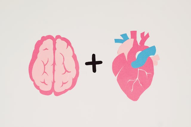
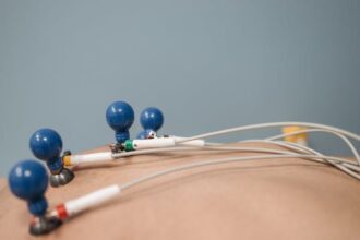
A recent investigation led by researchers at Brigham and Women’s Hospital, a prominent member of the Mass General Brigham healthcare system, indicates that heart rate could serve as a valuable indicator for determining optimal brain stimulation sites in individuals with depressive disorders when conventional brain imaging techniques are unavailable.
The study, featured in Nature Mental Health, examined data from 14 individuals exhibiting no symptoms of depression. By utilising transcranial magnetic stimulation (TMS) to target specific brain regions associated with depression, researchers observed concurrent effects on heart rate. This suggests that clinicians may identify and target relevant brain areas without the necessity of widely inaccessible brain scans.
Senior author Dr Shan Siddiqi of Brigham’s Department of Psychiatry and the Center for Brain Circuit Therapeutics explained that the concept emerged during a conference where Dutch researchers presented data on heart-brain coupling. The findings indicated the transient reduction of heart rate through TMS and the significance of the stimulation site. Dr. Siddiqi expressed enthusiasm for providing easier access to precision-targeted depression treatment worldwide, leveraging advanced technology in Boston.
Collaborating with colleagues at the Center for Brain Circuit Therapeutics, lead author Eva Dijkstra, MSc, integrated their expertise on heart-brain coupling with Brigham’s research on brain circuitry. This collaborative effort led to the identification of ten brain spots in each participant, categorised as either optimal (‘connected areas’) or non-optimal for depression treatment, with subsequent observation of heart rate fluctuations upon stimulation of each spot.
Dijkstra highlighted the promising discovery, emphasising the substantial heart-brain coupling observed in the connected areas for most participants. This finding holds the potential for customising TMS therapy based on individual brain targets, a development that could significantly improve the lives of individuals with depressive disorders, without the prerequisite of an MRI scan.
Dr. Siddiqi underscored the study’s broader implications, foreseeing its relevance in developing treatments applicable to cardiologists and emergency physicians in clinical settings. However, the study’s limitations lie in its small sample size and the inability to stimulate all possible brain spots.
Moving forward, the team is optimistic about refining their understanding of brain stimulation sites to ensure consistent changes in heart rate. Meanwhile, Dijkstra’s team in the Netherlands is conducting a larger study involving 150 individuals with treatment-resistant depression, with data analysis expected to significantly advance the research’s clinical translation later this year.
More information: Eva S. A. Dijkstra et al, Probing prefrontal-sgACC connectivity using TMS-induced heart–brain coupling, Nature Mental Health. DOI: 10.1038/s44220-024-00248-8
Journal information: Nature Mental Health Provided by Brigham and Women’s Hospital


