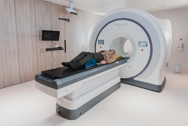
A study recently published in the journal Radiology by the Radiological Society of North America (RSNA) reveals that MRI images illustrating the brain’s structural and functional layout can forecast the progression of brain atrophy in individuals diagnosed with early-stage, mild Parkinson’s disease.
Parkinson’s disease is a neurodegenerative disorder identified by symptoms such as tremors, slow movements, or stiffness. These symptoms tend to worsen over time and may expand to include cognitive decline and sleep disturbances. Over 8.5 million people globally suffer from this condition, which has doubled over the last quarter-century, data from the World Health Organization (WHO) indicates.
A notable hallmark of Parkinson’s disease is the accumulation of abnormal alpha-synuclein protein variants within the brain. Under normal conditions, this protein exists benignly in the brain. Still, malfunctions in Parkinson’s misfold into aggregates that gather inside nerve cells, forming what are known as Lewy bodies and Lewy neurites. These aggregates further propagate throughout the brain, impairing nerve function.
The research team sought to determine whether visualising the structural and functional networks across the brain, using connectome maps derived from MRI scans, could predict the pattern of atrophy progression in patients with mild Parkinson’s disease. To this end, they analysed MRI data from 86 patients with mild Parkinson’s and 60 healthy controls to create these connectomes, subsequently developing an index to measure disease exposure.
The study found significant correlations between disease exposure at one and two years and brain atrophy at two and three years post-initial assessment. Models that included disease exposure effectively forecasted the accumulation of grey matter atrophy over three years in several brain regions.
Dr Federica Agosta, M.D., PhD, an associate professor of neurology at the Neuroimaging Research Unit of IRCCS San Raffaele Scientific Institute in Milan, Italy, and co-author of the study, remarked on the significance of these findings. She highlighted that the brain’s connectome, encompassing structural and functional aspects, demonstrated potential in predicting the progression of grey matter alterations in patients with mild Parkinson’s disease.
Dr Agosta further explained that neuron deterioration and the build-up of abnormal proteins could interfere with neural connections, hindering the transmission of neural signals and information integration across different brain areas. The study underscores the importance of MRI in intervention trials aimed at halting or slowing down the progression of the disease, particularly when tailored to individual patient data.
As the progression of Parkinson’s disease varies among patients, future research models should account for individual-specific starting conditions and incorporate personalised data to enhance their predictive accuracy. Dr. Agosta concluded by stressing the critical goal in neuroscience of understanding the brain’s network dynamics through the study of the human connectome, aspiring to identify new biomarkers that could influence the progression of Parkinson’s disease.
More information: Silvia Basaia et al, Brain Connectivity Networks Constructed Using MRI for Predicting Patterns of Atrophy Progression in Parkinson Disease, Radiology. DOI: 10.1148/radiol.232454
Journal information: Radiology Provided by Radiological Society of North America


