An artificial intelligence (AI) model that integrates MRI scans, biochemical tests, and clinical information has shown early promise in predicting whether a patient’s knee osteoarthritis will worsen. The study, led by Ting Wang of Chongqing Medical University in China, was published on August 21st in the open-access journal PLOS Medicine. The research suggests that this kind of integrated model may support clinicians in making earlier and more accurate treatment decisions for patients at risk of disease progression.
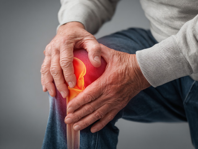
Knee osteoarthritis is a common and debilitating condition in which cartilage in the knee joint deteriorates over time, causing persistent pain and stiffness. Globally, it affects more than 300 million people and is a leading cause of disability among older adults. In severe cases, the disease often results in the need for total knee replacement surgery. Predicting which patients are most likely to experience worsening symptoms or structural decline could allow doctors to deliver more timely interventions, potentially slowing disease progression and improving quality of life.
Previous studies have suggested that predictive models may be strengthened by drawing upon multiple forms of patient data. For instance, MRI scans can reveal subtle changes in cartilage and joint space, while biochemical tests in blood or urine may capture metabolic or inflammatory signals. Clinical data, meanwhile, provides essential context about pain, mobility, and overall health. Yet, despite the promise of such multimodal approaches, few studies have successfully integrated all three forms of information into a single AI-driven model.
To address this gap, Wang and colleagues analysed data from 594 individuals with knee osteoarthritis, drawn from the Foundation of the National Institutes of Health Osteoarthritis Biomarkers Consortium. This included clinical assessments, biochemical test results, and a total of 1,753 MRI scans gathered over two years. The team used half the dataset to build a predictive model they called the Load-Bearing Tissue Radiomic plus Biochemical biomarker and Clinical variable Model (LBTRBC-M), and then validated its performance using the other half. The results showed that LBTRBC-M could accurately distinguish between patients who would experience worsening pain, combined pain and joint space narrowing, structural worsening without pain, or no decline at all over the following two years.
Importantly, the model was also tested in a clinical context by seven resident physicians. When these doctors used the LBTRBC-M system to guide their own predictions, their accuracy improved significantly—from just under 47 per cent to more than 65 per cent. This finding demonstrates the potential of AI models not only as research tools but as practical supports for medical decision-making. By synthesising complex data into actionable predictions, such systems could provide clinicians with an evidence-based means of identifying patients most at risk of rapid disease progression.
The authors emphasised that further refinement and broader validation are needed before LBTRBC-M can be implemented in routine clinical practice. Nevertheless, they underscored the potential of combining deep learning with longitudinal imaging and biochemical biomarkers to advance personalised care. “Our study shows that integrating these approaches significantly improves prediction of knee osteoarthritis progression, creating opportunities for earlier and more tailored interventions,” they wrote. Co-author Professor Changhai Ding added that the work highlights how AI can extract clinically meaningful insights from complex musculoskeletal datasets, paving the way for innovations that bridge technology and patient-centred care.
More information: Ting Wang et al, Predicting knee osteoarthritis progression using neural network with longitudinal MRI radiomics, and biochemical biomarkers: A modeling study, PLOS Medicine. DOI: 10.1371/journal.pmed.1004665
Journal information: PLOS Medicine Provided by PLOS