Researchers at the Mark and Mary Stevens Neuroimaging and Informatics Institute (Stevens INI), part of the Keck School of Medicine at the University of Southern California (USC), have introduced a pioneering brain imaging technique that uncovers how minute blood vessels in the brain pulse in rhythm with each heartbeat. These microscopic movements, invisible to conventional imaging, may hold crucial clues about how ageing affects the brain and could shed light on the mechanisms driving neurodegenerative conditions such as Alzheimer’s disease. The breakthrough, published in Nature Cardiovascular Research, represents a significant leap in understanding how vascular health shapes cognitive ageing, offering a window into processes that were previously hidden from human observation.
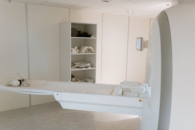
The USC team has developed the first noninvasive method capable of measuring what they term “microvascular volumetric pulsatility”—the rhythmic expansion and contraction of the brain’s tiniest vessels as blood surges through them. Using an ultra-high-field 7T magnetic resonance imaging (MRI) scanner, the researchers demonstrated that microvascular pulsations become more pronounced with age, particularly in the brain’s deep white matter. This region, responsible for facilitating communication between different brain areas, is especially vulnerable to diminished blood flow from distal arteries—vessels that carry oxygenated blood into the brain’s most remote tissues. According to the study, increasing microvessel pulsatility may contribute to the gradual deterioration of the brain’s vascular systems, which could, in turn, accelerate memory decline and cognitive impairment in older adults.
“Arterial pulsation acts as the brain’s own pump, promoting the circulation of fluids and the clearance of waste,” explained senior author Danny JJ Wang, PhD, professor of neurology and radiology at the Keck School of Medicine. “Our new imaging method allows us to see for the first time, in living people, how the volume of these tiny blood vessels fluctuates with age and vascular risk factors. This discovery provides a vital new tool for exploring how changes in microvascular function contribute to dementia, small vessel disease, and overall brain health.” For decades, scientists have understood that stiffness and abnormal pulsatility in large arteries increase the risk of conditions such as stroke and vascular dementia. Yet, until now, there has been no safe, non-invasive way to study similar dynamics in the brain’s smallest vessels, which has limited human research to indirect inferences or animal models.
To overcome these barriers, the USC researchers combined two sophisticated MRI techniques—vascular space occupancy (VASO) and arterial spin labelling (ASL)—to track minute fluctuations in vessel volume over the cardiac cycle. This integration enabled them to visualise subtle variations in microvascular pulsation across different regions of the brain. Their results revealed that older participants exhibited significantly higher levels of microvascular pulsatility than younger adults, particularly within deep white matter regions. Moreover, hypertension amplified these changes, suggesting that elevated blood pressure can exacerbate age-related vascular alterations. “Our findings establish a missing link between the macro-level vascular changes visible in large artery imaging and the microvascular damage observed in ageing and Alzheimer’s disease,” noted lead author Fanhua Guo, PhD, a postdoctoral researcher in Wang’s laboratory.
The implications of these findings extend beyond vascular mechanics. The researchers believe that excessive pulsatility in small vessels may impair the function of the brain’s recently discovered “glymphatic system”—a network that clears metabolic waste, including beta-amyloid, a protein associated with Alzheimer’s pathology. Disruption to this cleansing system could hinder the removal of harmful waste products, leading to their accumulation over time and thereby hastening neurodegeneration. “The ability to measure these microscopic vascular pulses in vivo marks a transformative step forward,” said Arthur W. Toga, PhD, director of the Stevens INI. “This technology not only deepens our understanding of brain ageing but could also pave the way for early diagnosis and more accurate monitoring of neurodegenerative diseases.”
Looking ahead, the team is working to adapt the technique for broader clinical use, including its application on standard 3T MRI scanners, which are far more accessible than the research-focused 7T systems. They also plan to investigate whether microvascular volumetric pulsatility could serve as a predictive biomarker for cognitive decline, potentially identifying individuals at risk of dementia before symptoms appear. “This is only the beginning,” Wang remarked. “Our long-term vision is to bring this technology from the research laboratory into everyday clinical practice, where it can guide diagnosis, prevention, and treatment for millions facing age-related neurological diseases. By revealing the hidden rhythms of the brain’s smallest blood vessels, we are uncovering an entirely new dimension of human physiology—one that may redefine how we understand, preserve, and restore brain health throughout the lifespan.”
More information: Fanhua Guo et al, Assessing cerebral microvascular volumetric with high-resolution 4D cerebral blood volume MRI at 7 T, Nature Cardiovascular Research. DOI: 10.1038/s44161-025-00722-1
Journal information: Nature Cardiovascular Research Provided by Keck School of Medicine of USC


